
- Title
-
Tdrd12 Is Essential for Germ Cell Development and Maintenance in Zebrafish
- Authors
- Dai, X., Shu, Y., Lou, Q., Tian, Q., Zhai, G., Song, J., Lu, S., Yu, H., He, J., Yin, Z.
- Source
- Full text @ Int. J. Mol. Sci.

ZFIN is incorporating published figure images and captions as part of an ongoing project. Figures from some publications have not yet been curated, or are not available for display because of copyright restrictions. PHENOTYPE:
|
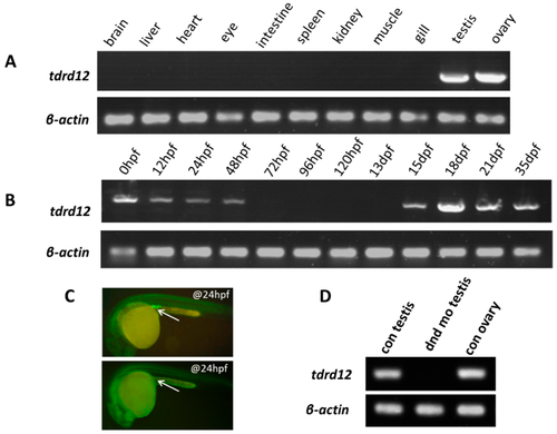
Maternal and germ cell-related expression patterns of tdrd12. (A) Tissue distribution of endogenous tdrd12 transcripts with RT-PCR indicates its gonad-specific patterns in adult zebrafish; (B) The presence of endogenous tdrd12 transcripts at different developmental stages reveals its maternal expression pattern in unfertilized eggs. The expression of zebrafish tdrd12 is decreased in the early embryonic stage until the 48 hpf stage. The tdrd12 transcripts reappear from 15 dpf to the adult stage in the gonads; (C) Presence of PGCs visualized with injected EGFP-dnd 3’UTR mRNA in control larva (upper panel, injected with control morpholino) and PGC-depleted larva (lower panel, injected with dnd morpholino) at the 24-hpf stage. The arrowheads here show the presence of zebrafish PGCs in larva; and (D) Presence of tdrd12 transcripts detected in adult gonadal tissues of the control fish and PGC-depleted dnd morphants (no ovary). The transcripts of β-actin2 were amplified from the same templates as an internal control to check the quality of the cDNA. |
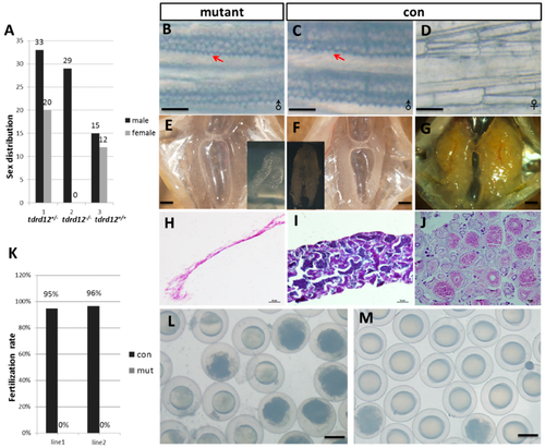
Tdrd12 deficiency results in masculinization and infertility in zebrafish. (A) Sex ratios of the progenies from the in-cross of tdrd12 heterozygous mutant fish (90 dpf). Among the progenies, all homozygous offspring developed into males. The total number of progenies for this analysis is 109. tdrd12+/−: heterozygous progenies; tdrd12+/+: wild-type progenies; tdrd12−/−: homozygous progenies; (B–D) Appearance of the breeding tubercle (BT) clusters (red arrowheads) in the pectoral fin of all homozygous mutant fish (B) and wild-type males (C), but not in wild-type females (D), scale bar = 250 μm; (E–G) Anatomical views of the gonadal tissues of the Tdrd12-deficient fish and wild-type adults. Only atrophied testes were observed in Tdrd12-deficient adults (E), while normal testes (F) and ovary (G) could be observed in wild-types adults, scale bar = 500 μm; (H–J) Histological analyses of the gonadal tissues indicate no signs of germ cells in the atrophied testes of the Tdrd12-deficient adults (H), while normal spermatogenesis (I) and oogenesis (J) progresses in the wild-type adults, scale bar = 50 μm; (K) The fertilization rates of the mating between wild-type (tdrd12+/+) males and Tdrd12-deficient fish with wild-type females at the 120-dpf stage were recorded. The average fertilization rates of the 10 fish from three separate experiments were measured. Each group consists of 10 pairs of fish. The data shown here represent the means ± standard error of the mean; and (L,M) Morphological observations of embryonic development at 11 hpf derived from the mating between wild-type females with Tdrd12-deficient adults (L) and wild-type males (M); eggs from wild-type females could be induced by Tdrd12-deficient fish, but no successful fertilization could be found, scale bar = 250 μm. |
|
ZFIN is incorporating published figure images and captions as part of an ongoing project. Figures from some publications have not yet been curated, or are not available for display because of copyright restrictions. |
|
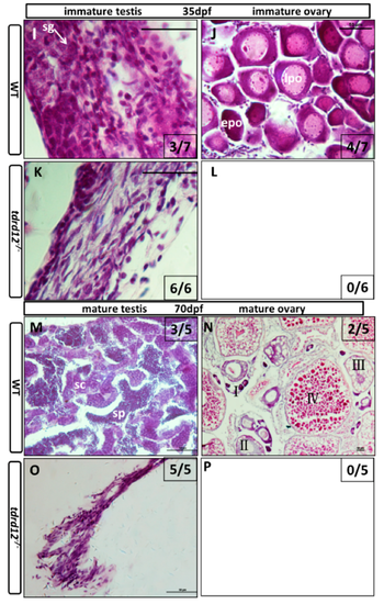
Masculinization occurred in Tdrd12-deficient zebrafish during the late “juvenile ovary” stage. Histological features with hematoxylin-eosin staining of cryo-sections were assessed from gonadal tissues in Tdrd12-deficient zebrafish and their wild-type siblings at various stages. (A–D) At the 18-dpf stage, typical stage Ia in the “juvenile ovary” was observed in all wild-type fish gonadal tissue (A, 7/7), and most Tdrd12-deficient fish gonadal tissue (C, 6/7). However, typical pyknotic cells (pc) in early testes could not been found in wild-type gonadal samples (B, 0/7) and only some of Tdrd12-deficient fish gonadal tissue samples (D, 1/7); (E–H) At the 25-dpf stage, typical pyknotic cells (pc) and spermatogonia (sg) in early testes were observed in some of the wild-type fish gonadal tissue (E, 2/7) and all Tdrd12-deficient fish gonadal tissues were examined (G, 6/6). However, the typical epo stage ((“early” perinucleolar oocytes): early stage IB) in early ovaries could be found in some of the wild-type gonadal tissue samples (F, 5/7). No ovary-like gonads could be found in Tdrd12-deficient gonadal samples (H); (I–L) At 35 dpf, typical spermatogonium (I) or oogonium (J) could be seen in the gonadal samples of the wild-type males (I, 3/7) and females (J, 4/7). However, only the filament-like testes with some Sertoli-like and Leydig-like cells, but no spermatogonium-like cells, were seen in the gonadal samples of the Tdrd12-deficient fish (K, 6/6). No ovary-like tissue was found in mutant fish (L, 0/6); (M–P). At 70 dpf, various spermatogonium (I) or oogonium (J) stages could be seen in the gonadal samples of the wild-type males (M, 3/5) and females (N, 2/5). However, only the filament-like testes without any sign of the presence of germ cells could be seen in all the gonadal samples of Tdrd12-deficient fish (O, 5/5). No ovary-like tissue has been found in mutant fish (L, 0/5). The gonadal tissues in (A–H) are circled with a black dotted line, and the white arrowheads indicate typical cell types in each pictures. (A–H,I,K): 1000×; (J,M,O): 400×; and (N): 100×. Scale bar for all pictures represents 50 μm. Pyknotic cells (pc), spermatogonia (sg), sperm (sp), spermatocytes (sc); stages of oogenesis: stage I, stage I is divided into stage Ia and Ib. (epo (“early” perinucleolar oocytes): early stage of Ib); lpo (“late” perinucleolar oocytes): late stage of Ib), II, III, and IV. 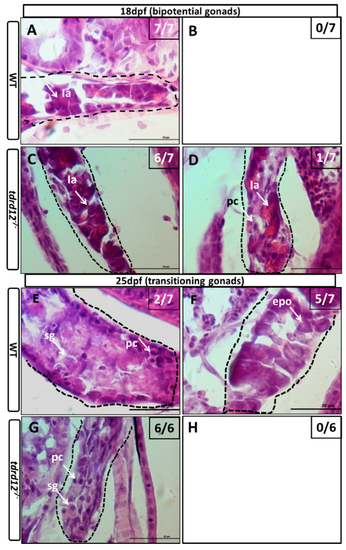
PHENOTYPE:
|
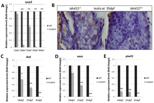
Meiotic defects and failure of germ cell maintenance in Tdrd12-deficient zebrafish. (A) Normalized expression of the meiotic recombination-specific gene sycp3 (synaptonemal complex protein 3) in the developing gonads were examined in gonadal tissue samples in Tdrd12-deficient fish and their wild-type siblings at 13, 18, 25, and 35 dpf; (B) In situ cell death in the immature testis of tdrd12+/+ and tdrd12−/− at 35 dpf, with abundant signals (brown) in the Tdrd12-deficient testis. Normalized expression of the germ cell-specific genes dnd (C), vasa (D), and piwil1 (E) in the developing gonads were examined in gonadal tissue samples in Tdrd12-deficient fish and their wild-type siblings at 13, 18, 25, and 35 dpf. The tails of the larvae were collected for genotyping, while the rest of the body was used for total RNA isolation. The numbers of the examined fish for the assays at the 13, 18, 25, and 35 dpf stages for each genotype were 25, 20, 12, and 10, respectively. The experiments were performed with three biological repeats. * indicates the difference at p < 0.05 vs. wild type; ** indicates the significant difference at p < 0.01 vs. wild type. β-actin2 was selected as the most suitable and invariant reference gene for our samples from gapdh, β-actin2, and ef1a testing according to the published reports [33,34]. PHENOTYPE:
|
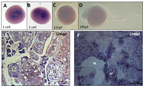
Expression pattern of tdrd12 in the germ cells. In situ hybridization of the tdrd12 probe in embryos at the 1- to 2-cell stage (A and B), 11 hpf (C), 24 hpf (D), and cryo-sections of adult gonads are present in the ovary (E) and testis (F). The types and stages of the cells were labeled, and the signals are indicated in purple. However, the signals vanished at 11 hpf and 24 hpf (C and D). |
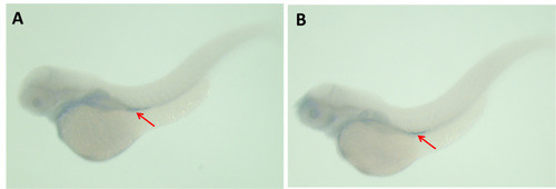
Normal development of the PGCs seen in tdrd12-deficient larvae from heterozygous parents. (A,B) PGCs visualized with the vasa riboprobe in wild-type larvae (A) and tdrd12-deficient larvae from the heterozygous parents (B) at 24 hpf. PGCs are indicated with arrowheads. EXPRESSION / LABELING:
|
|
ZFIN is incorporating published figure images and captions as part of an ongoing project. Figures from some publications have not yet been curated, or are not available for display because of copyright restrictions. |