
Image
|
Figure Caption
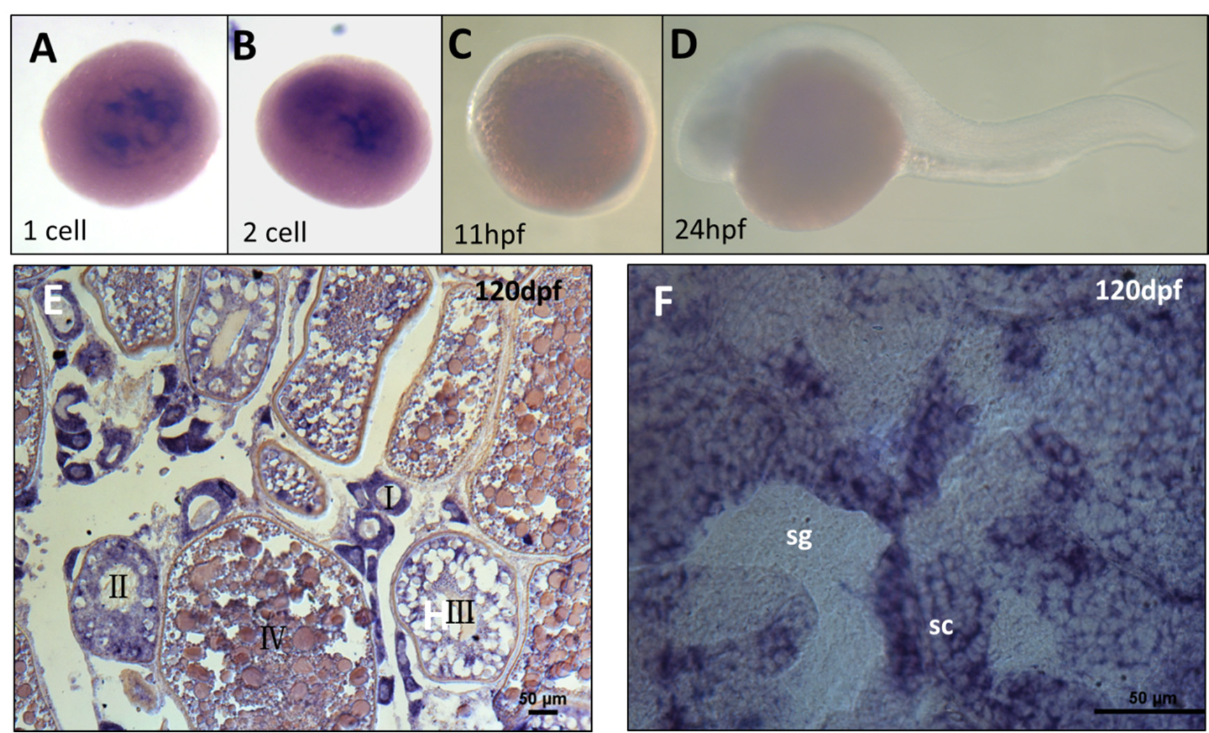
Fig. S1
Expression pattern of tdrd12 in the germ cells. In situ hybridization of the tdrd12 probe in embryos at the 1- to 2-cell stage (A and B), 11 hpf (C), 24 hpf (D), and cryo-sections of adult gonads are present in the ovary (E) and testis (F). The types and stages of the cells were labeled, and the signals are indicated in purple. However, the signals vanished at 11 hpf and 24 hpf (C and D).
Figure Data
Acknowledgments
This image is the copyrighted work of the attributed author or publisher, and
ZFIN has permission only to display this image to its users.
Additional permissions should be obtained from the applicable author or publisher of the image.
Full text @ Int. J. Mol. Sci.