
Fig. 6
|
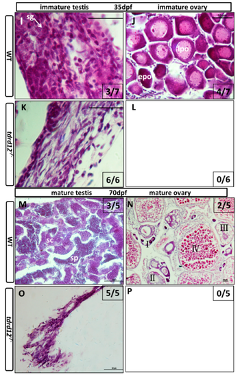
Masculinization occurred in Tdrd12-deficient zebrafish during the late “juvenile ovary” stage. Histological features with hematoxylin-eosin staining of cryo-sections were assessed from gonadal tissues in Tdrd12-deficient zebrafish and their wild-type siblings at various stages. (A–D) At the 18-dpf stage, typical stage Ia in the “juvenile ovary” was observed in all wild-type fish gonadal tissue (A, 7/7), and most Tdrd12-deficient fish gonadal tissue (C, 6/7). However, typical pyknotic cells (pc) in early testes could not been found in wild-type gonadal samples (B, 0/7) and only some of Tdrd12-deficient fish gonadal tissue samples (D, 1/7); (E–H) At the 25-dpf stage, typical pyknotic cells (pc) and spermatogonia (sg) in early testes were observed in some of the wild-type fish gonadal tissue (E, 2/7) and all Tdrd12-deficient fish gonadal tissues were examined (G, 6/6). However, the typical epo stage ((“early” perinucleolar oocytes): early stage IB) in early ovaries could be found in some of the wild-type gonadal tissue samples (F, 5/7). No ovary-like gonads could be found in Tdrd12-deficient gonadal samples (H); (I–L) At 35 dpf, typical spermatogonium (I) or oogonium (J) could be seen in the gonadal samples of the wild-type males (I, 3/7) and females (J, 4/7). However, only the filament-like testes with some Sertoli-like and Leydig-like cells, but no spermatogonium-like cells, were seen in the gonadal samples of the Tdrd12-deficient fish (K, 6/6). No ovary-like tissue was found in mutant fish (L, 0/6); (M–P). At 70 dpf, various spermatogonium (I) or oogonium (J) stages could be seen in the gonadal samples of the wild-type males (M, 3/5) and females (N, 2/5). However, only the filament-like testes without any sign of the presence of germ cells could be seen in all the gonadal samples of Tdrd12-deficient fish (O, 5/5). No ovary-like tissue has been found in mutant fish (L, 0/5). The gonadal tissues in (A–H) are circled with a black dotted line, and the white arrowheads indicate typical cell types in each pictures. (A–H,I,K): 1000×; (J,M,O): 400×; and (N): 100×. Scale bar for all pictures represents 50 μm. Pyknotic cells (pc), spermatogonia (sg), sperm (sp), spermatocytes (sc); stages of oogenesis: stage I, stage I is divided into stage Ia and Ib. (epo (“early” perinucleolar oocytes): early stage of Ib); lpo (“late” perinucleolar oocytes): late stage of Ib), II, III, and IV. 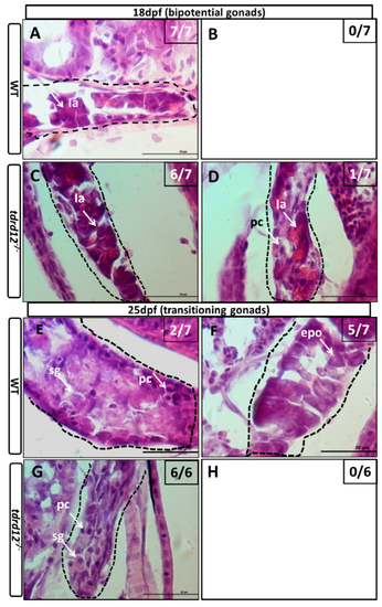
|
| Fish: | |
|---|---|
| Observed In: | |
| Stage Range: | Days 14-20 to Days 45-89 |