
- Title
-
Ablation of EYS in zebrafish causes mislocalisation of outer segment proteins, F-actin disruption and cone-rod dystrophy
- Authors
- Lu, Z., Hu, X., Liu, F., Soares, D.C., Liu, X., Yu, S., Gao, M., Han, S., Qin, Y., Li, C., Jiang, T., Luo, D., Guo, A.Y., Tang, Z., Liu, M.
- Source
- Full text @ Sci. Rep.

ZFIN is incorporating published figure images and captions as part of an ongoing project. Figures from some publications have not yet been curated, or are not available for display because of copyright restrictions. |
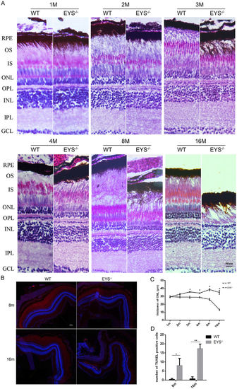
The thickness of ONL decreased and the apoptosis of retinal cells increased in the EYS−/− zebrafish. (A) Retinal histology analysis of WT and EYS−/− zebrafish. Retinal sections of EYS−/− zebrafish stained with hematoxylin/eosin (HE) at indicated ages. RPE, retinal pigment epithelium; OS, outer segment; IS, inner segment; ONL, outer nuclear layer; OPL, outer plexiform layer; INL, inner nuclear layer; IPL, inner plexiform layer; GCL, ganglion cell layer. Scale bars: 50 μm. (B) TUNEL staining between WT and EYS−/− zebrafish. Scale bars: 50 μm. (C) Comparison of the ONL thickness between WT and EYS−/− zebrafish at indicated ages (n = 3). Quantitative analysis revealed a significant reduction of the ONL thickness at 3 mpf (P = 0.0479), 4 mpf (P = 0.0258), 8 mpf (P = 0.0122), 16 mpf (P = 0.0272) in the EYS−/− zebrafish. (D) Quantification of TUNEL positive cells in WT and EYS−/− zebrafish at indicated ages (n = 3). Quantitative analysis revealed a significant increase of TUNEL positive cells at 8 mpf (P = 0.0341), 16 mpf (P = 0.0028) in the EYS−/− zebrafish. PHENOTYPE:
|

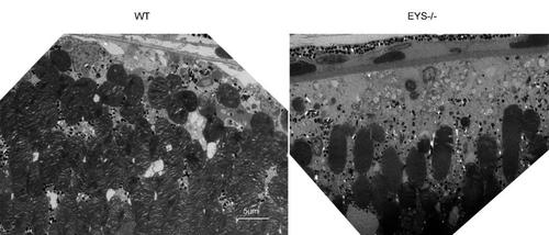
Ultrastructural analysis of the WT and EYS−/− zebrafish photoreceptors at 3 mpf. The morphological and disk-stacking of OS is normal between the WT and EYS−/− zebrafish (A,B). The number of photoreceptors in EYS−/− zebrafish is less than WT zebrafish (C,D). Scale bars: 1 μm in (A,B); 5 μm in (C,D). PHENOTYPE:
|
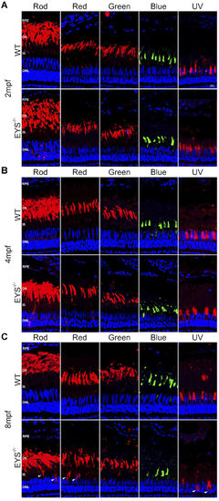
Photoreceptor outer segment is affected in EYS−/− zebrafish. Retinal cryosections from WT and EYS−/− zebrafish were labelled rods and cones (red, green, blue and UV cone) with specific antibodies at the ages of 2 (A), 4(B) and 8(C) mpf. White arrows indicate the mislocalised OS proteins. Scale bars: 10 μm. PHENOTYPE:
|
|
ZFIN is incorporating published figure images and captions as part of an ongoing project. Figures from some publications have not yet been curated, or are not available for display because of copyright restrictions. PHENOTYPE:
|
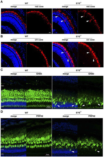
Photoreceptor OS proteins were mislocalised in EYS−/− zebrafish. (A) Retinal cryosections from WT and EYS−/− zebrafish were immunostained with anti-opn1LW antibodies at 10 dpf. White arrows indicated the mislocalised opn1LW proteins. Scale bars: 10 μm. (B) Retinal cryosections from WT and EYS−/− zebrafish were immunostained with anti-opn1SW1 antibodies at 10 dpf. White arrows indicated the mislocalised opn1SW1 proteins. Scale bars: 10 μm. (C) Retinal cryosections from WT and EYS−/− zebrafish were immunostained with anti-GNB3 antibodies at the age of 5 mpf. White arrows indicate the mislocalised GNB3 proteins. Scale bars: 50 μm. (D) Retinal cryosections from WT and EYS−/− zebrafish were immunostained with anti-PRPH2 antibodies at the age of 7 mpf. White arrows indicated the mislocalised PRPH2 proteins. Scale bars: 50 μm. |

F-actin disruption in EYS−/− zebrafish. Retinal cryosections were immunostained with phalloidin between WT and EYS−/− zebrafish. (A) F-actin was disrupted, slightly, in EYS−/− zebrafish at 2 mpf. (B) F-actin was disrupted, substantially, in EYS−/− zebrafish at 7 mpf. White arrows indicate the disrupted F-actin. Scale bars: 20 μm. PHENOTYPE:
|
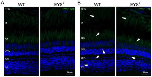
Immunostaining of the WT and EYS-/- zebrafish retinas at 3mpf using the EYS antibody from Novus Biological (Cat# NBP1-90038, which is used in the reported open bio. paper) (1:300) A and (1:100) B.White arrows indicate the possible positive signal, it was not only found between the retinal pigment epithelium (RPE) and ONL but also located in the ONL and inner nuclear layer (INL). Scale bars: 20μm. |

The whole retina sections with HE staining at the age of 16mpf. Scale bars: 100μm. PHENOTYPE:
|

TUNEL analysis of WT and EYS-/- zebrafish retina at 2mpf. Scale bars: 50μm. PHENOTYPE:
|
Retinal ultrastructural of WT and EYS-/- zebrafish at 10mpf. Scale bars: 5μm. |

The whole retina sections of immunofluorescence analysis with different opsin antibodies at the age of 16mpf. Scale bars: 50μm. |
|
ZFIN is incorporating published figure images and captions as part of an ongoing project. Figures from some publications have not yet been curated, or are not available for display because of copyright restrictions. |

Immunostaining of the WT and EYS-/- zebrafish retinas at 10dpf with phalloidin. |