- Title
-
Contact-Mediated Inhibition Between Oligodendrocyte Progenitor Cells and Motor Exit Point Glia Establishes the Spinal Cord Transition Zone
- Authors
- Smith, C.J., Morris, A.D., Welsh, T.G., Kucenas, S.
- Source
- Full text @ PLoS Biol.
|
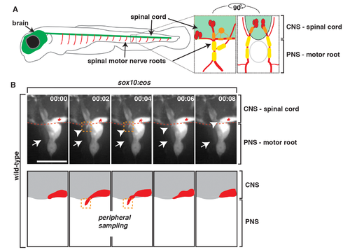
PNS glia restrict OPC membrane processes in the PNS. (A) Schematic of zebrafish spinal motor roots showing the CNS (green) consisting of the spinal cord and brain and the PNS (red). Insets show zoomed representations of a single spinal motor nerve root with motor axons (red), OPCs (orange), and PNS glia (yellow). Lateral and cross-section views are shown. (B) Frames captured from a 14-h time-lapse video beginning at 58 hpf in a Tg(sox10:eos) embryo. Numbers in upper right corners denote time lapsed from the first frame of the figure. (A) At approximately 68 hpf (00:00), peripheral glial cells (arrow) and OPCs (asterisk) can be seen. Just 2 min later (00:02), an OPC process (arrowhead/orange box) extended out of the spinal cord, contacted the motor root glial cell (arrow), and quickly retracted, all within 6 min. Traced schematic below shows spinal cord (grey) and OPC (red), at the corresponding times. All images are lateral views of the motor and sensory root with dorsal to the top and anterior to the left. Scale bar, 15 µm. EXPRESSION / LABELING:
|
|
sox10+ motor root glial cells originate in the CNS. A 90-degree rotation of images captured from a 24-h time-lapse video beginning at 48 hpf in a Tg(sox10:eos) embryo. Numbers in lower right corners denote time lapsed from the first frame of the figure. At approximately 56 hpf (00:00), a sox10+ cell (asterisk) migrated from the spinal cord, pinched through the MEP (arrow), and migrated into the periphery. Migration path shows the migration of neural crest versus CNS-derived sox10+ cells during this developmental stage. Dashed line marks lateral edge of the spinal cord. Scale bar, 25 µm. EXPRESSION / LABELING:
|
|
Motor root-associated glia are distinct from neural crest-derived glia. (A) In a Tg(sox10:eos) embryo exposed to UV light at 48 hpf and imaged at 80 hpf, photoconverted cells (red, arrowhead) are only observed along sensory axons while unconverted cells (green, arrow) populate the motor root. CNS and PNS portions are denoted in brackets. (B) Quantification of photoconversion experiments shows CNS-derived, unconverted sox10+ cells develop between 48 and 72 hpf, whereas converted sox10+ cells that generate the DRG are present before 48 hpf (30 nerves scored). (C) Frames captured from a 24-h time-lapse video beginning at 48 hpf in a Tg(sox10:eos) embryo that was exposed to UV light at 48 hpf and DRG ablation immediately after. Numbers in upper right corners denote stage of development. At 51.3 hpf (03:20), a CNS-derived sox10+ cell (green, arrow) associated with motor axons and generated motor root glial cells even in the presence of dying DRG cells (red). All images are lateral views of the motor and sensory root with dorsal to the top and anterior to the left. CNS and PNS portions are denoted in brackets. Scale bars, 25 µm. EXPRESSION / LABELING:
|
|
sox10+ motor root glia arise from olig2+ precursors in the spinal cord. (A) Frames captured from a 24-h time-lapse video of a Tg(olig2:dsred) larvae showing olig2+ cells (arrow) migrating out of the CNS, pinching at the MEP, and associating with the spinal motor root axons. Numbers in lower right corners denote time lapsed from the first frame of the figure. Brackets show the CNS and PNS portions in the images. (B) In Tg(sox10:eos) embryos injected with a nkx2.2a MO, CNS-derived sox10+ motor glial cells (arrow) are indistinguishable from wild-type nerves. In contrast, in olig2 MO-injected embryos, motor root glial cells are absent. In both instances, sensory glial cells are unaffected (arrowheads). (C) Quantification of data from panel B (>50 nerves were scored for each perturbation). (D) In a Tg(sox10:eos);Tg(olig2:dsred) embryo at 56 hpf, a sox10+ cell located on the motor nerve root expresses olig2 and sox10 (open arrowhead). Neighboring sox10+ cells in the DRG (asterisk) do not express olig2. (E) In a Tg(nkx2.2a:megfp);Tg(olig2:dsred) embryo at 72 hpf, nkx2.2+ perineurial glia are distinct from olig2+ MEP glia (open arrowhead). All images are lateral views of the motor and sensory root with dorsal to the top and anterior to the left. Scale bars, 25 µm. EXPRESSION / LABELING:
|
|
MEP glia express foxd3. Frames captured from a 24-h time-lapse video of a Gt(foxd3:mcherry) transgenic embryo showed a foxd3+ cell (arrow) located inside the spinal cord (dotted line), migrate ventrally, exit the spinal cord at the MEP, and associate with a spinal motor root axon. The neural crest-derived population (arrowhead) is posterior to the CNS-derived cell. (B) Frames captured from the above video of a Tg(gfap:egfp);Gt(foxd3:mcherry) embryo rotated 90 degrees showed a foxd3+ cell starts inside the spinal cord, as marked by gfap+ endfeet along the lateral edge of the cord, then migrates through the TZ, and associates with spinal root axons in the PNS. Scale bar, 25 µm. EXPRESSION / LABELING:
|
|
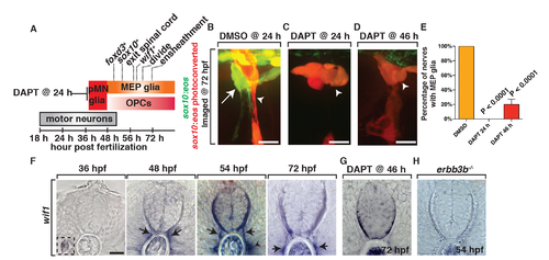
MEP glia originate from ventral spinal cord progenitors and express wif1. (A) Summary of MEP glial development and the timing of marker expression. olig2 is not included because the initiation of expression is difficult to determine with surrounding olig2+ cells already present during this stage of development. (B–D) In Tg(sox10:eos) embryos exposed to UV light at 48 hpf and imaged at 72 hpf, (B) DMSO-treated animals have MEP glia, whereas (C) embryos treated with DAPT at 24 hpf and (D) 46 hpf do not. (E) Quantification of data from panel D. (F) In situ hybridization with wif1 riboprobe at 36, 48, 54, and 72 hpf shows timing of wif1 expression at the root denoted by arrows. Arrowhead denotes the horizontal myoseptum staining. Inset at 36 hpf shows wif1+ staining is expressed at the lateral line, as has been previously described. (G–H) At 54 hpf in erbb3b (H) mutants and at 72 hpf in embryos treated with DAPT at 46 hpf (G), we observed an absence of wif1 staining along spinal motor roots. Scale bars, 25 µm. EXPRESSION / LABELING:
PHENOTYPE:
|
|
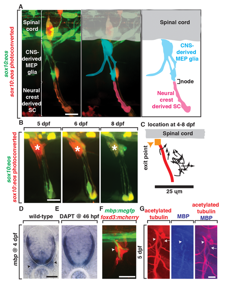
MEP glia and their descendents myelinate the spinal motor root. (A) In images from Tg(sox10:eos) animals exposed to UV at 48 hpf and imaged at 80 hpf, CNS-derived MEP glia ensheath the root and neural crest-derived Schwann cells (SCs) ensheath more distal portions of spinal nerves. CNS-derived MEP glia are overlayed with blue labeling and neural crest-derived SCs in pink. (B) In images of Tg(sox10:eos) embryos where single cells were photoconverted at 5 dpf and imaged on sequential days until 8 dpf, MEP glial derivatives remain ensheathed around spinal motor root axons. (C) Movement of single photoconverted MEP glia from 4–8 dpf. Orange box denotes the MEP, and red line denotes the motor axon. (D) In situ hybridization with mbp riboprobe showed mbp+ staining (denoted by arrowhead) at the motor root at 4 dpf is absent in (E) DAPT-treated animals that lack MEP glia. (F) In a Tg(mbp:megfp);Gt(foxd3:mcherry) embryo at 4 dpf, foxd3+ cells are located along a myelinated nerve (arrow). (G) In 5 dpf embryos stained with acetylated tubulin and MBP, MBP+ axons are present at the root where MEP glia reside. Scale bars, 25 µm. EXPRESSION / LABELING:
PHENOTYPE:
|
|
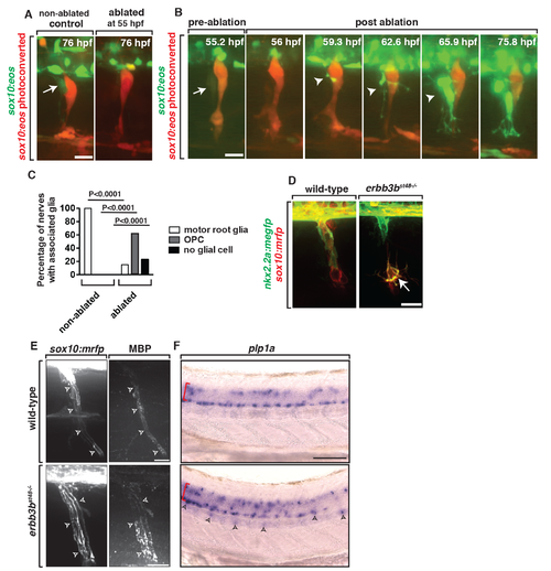
Ablation of MEP glia disrupts the MEP TZ. (A) In a control Tg(sox10:eos) embryo that was exposed to UV light at 48 hpf, motor (green) and sensory root (red) glial cells ensheath axons. When the CNS-derived MEP glial cell progenitor was ablated at 55 hpf and then imaged at 76 hpf, all sox10+ motor root glial cells were absent. (B) Frames captured from a 16-h time-lapse video beginning at 56 hpf in a Tg(sox10:eos) embryo exposed to UV light at 48 hpf. Numbers in upper right corners denote stage of embryos. When the MEP glial cell progenitor (arrow) was ablated at 55 hpf, OPC processes (arrowhead) extended out of the spinal cord before OPC cell bodies exited. All images are lateral views of the motor and sensory root with dorsal to the top and anterior to the left. (C) Quantification of the data in panel B (n = 46 nerves). (D) In Tg(nkx2.2a:mgfp);Tg(sox10:mrfp);erbb3b−/− embryos at 55 hpf, motor root glial cells are absent and OPCs (arrows) are in the PNS. (E) In Tg(sox10:mrfp) larva at 8 dpf stained with MBP, MBP+ cells were ensheathed around the root in wild-type and erbb3 animals. (F) In situ hybridization of plp1a in wild-type and erbb3b mutant showed OPCs in the PNS in erbb3b mutants myelinate with CNS myelin. Scale bars, 25 µm. |


