
Fig. 7
- ID
- ZDB-FIG-150128-8
- Publication
- Smith et al., 2014 - Contact-Mediated Inhibition Between Oligodendrocyte Progenitor Cells and Motor Exit Point Glia Establishes the Spinal Cord Transition Zone
- Other Figures
- All Figure Page
- Back to All Figure Page
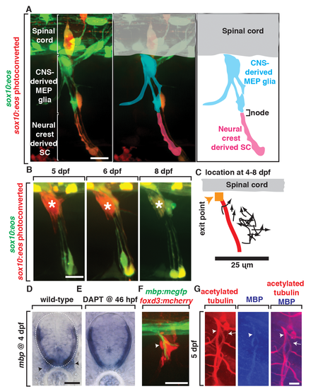
MEP glia and their descendents myelinate the spinal motor root. (A) In images from Tg(sox10:eos) animals exposed to UV at 48 hpf and imaged at 80 hpf, CNS-derived MEP glia ensheath the root and neural crest-derived Schwann cells (SCs) ensheath more distal portions of spinal nerves. CNS-derived MEP glia are overlayed with blue labeling and neural crest-derived SCs in pink. (B) In images of Tg(sox10:eos) embryos where single cells were photoconverted at 5 dpf and imaged on sequential days until 8 dpf, MEP glial derivatives remain ensheathed around spinal motor root axons. (C) Movement of single photoconverted MEP glia from 4–8 dpf. Orange box denotes the MEP, and red line denotes the motor axon. (D) In situ hybridization with mbp riboprobe showed mbp+ staining (denoted by arrowhead) at the motor root at 4 dpf is absent in (E) DAPT-treated animals that lack MEP glia. (F) In a Tg(mbp:megfp);Gt(foxd3:mcherry) embryo at 4 dpf, foxd3+ cells are located along a myelinated nerve (arrow). (G) In 5 dpf embryos stained with acetylated tubulin and MBP, MBP+ axons are present at the root where MEP glia reside. Scale bars, 25 µm. |
| Genes: | |
|---|---|
| Antibody: | |
| Fish: | |
| Conditions: | |
| Anatomical Terms: | |
| Stage Range: | Protruding-mouth to Days 7-13 |
| Fish: | |
|---|---|
| Condition: | |
| Observed In: | |
| Stage: | Day 4 |