
Fig. 5
- ID
- ZDB-IMAGE-161206-21
- Genes
- Publication
- Smith et al., 2014 - Contact-Mediated Inhibition Between Oligodendrocyte Progenitor Cells and Motor Exit Point Glia Establishes the Spinal Cord Transition Zone
- All Figures
- Figures for Smith et al., 2014
|
Fig. 5
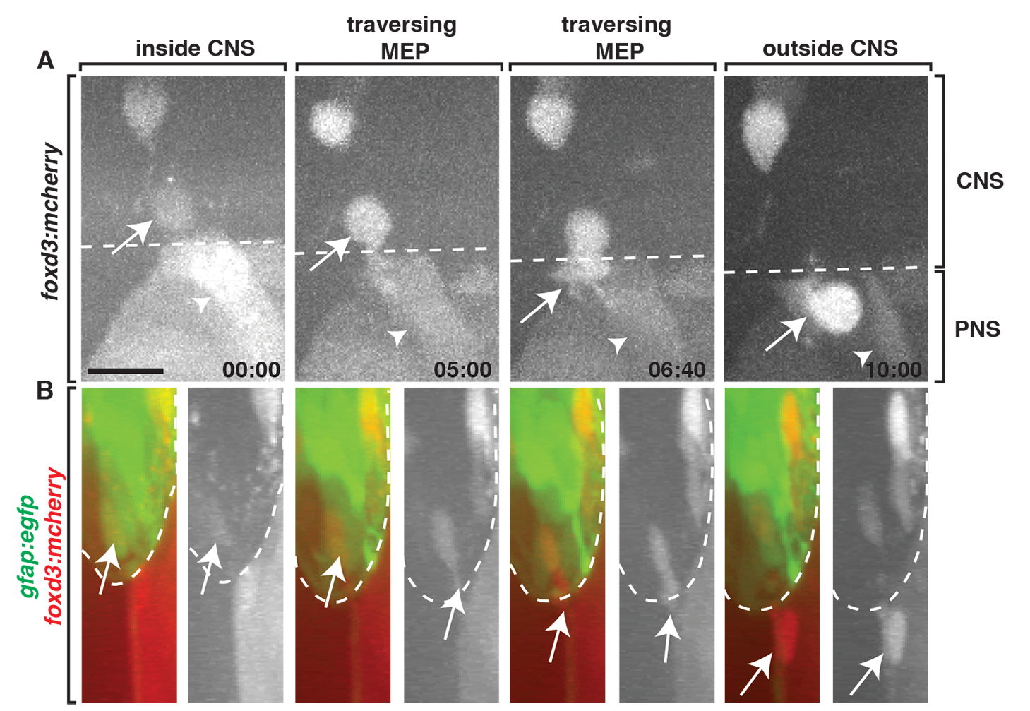
MEP glia express foxd3.
Frames captured from a 24-h time-lapse video of a Gt(foxd3:mcherry) transgenic embryo showed a foxd3+ cell (arrow) located inside the spinal cord (dotted line), migrate ventrally, exit the spinal cord at the MEP, and associate with a spinal motor root axon. The neural crest-derived population (arrowhead) is posterior to the CNS-derived cell. (B) Frames captured from the above video of a Tg(gfap:egfp);Gt(foxd3:mcherry) embryo rotated 90 degrees showed a foxd3+ cell starts inside the spinal cord, as marked by gfap+ endfeet along the lateral edge of the cord, then migrates through the TZ, and associates with spinal root axons in the PNS. Scale bar, 25 µm.