- Title
-
Duplication of the IGFBP-2 gene in teleost fish: protein structure and functionality conservation and gene expression divergence
- Authors
- Zhou, J., Li, W., Kamei, H., and Duan, C.
- Source
- Full text @ PLoS One
|
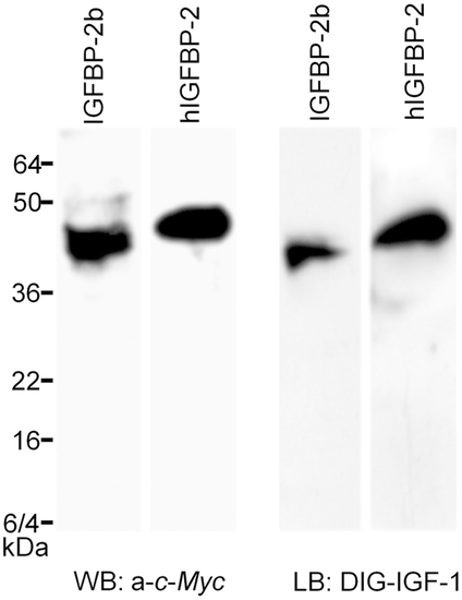
Zebrafish igfbp-2b encodes a secreted protein that binds IGFs. Conditioned media was prepared from HEK293T cells transfected with pCMV-human IGFBP-2-Myc (hIGFBP-2) or pCMV-zebrafish IGFBP-2b-Myc plasmids. The conditioned media were analyzed by Western immunoblotting (WB) using c-Myc antibody (left panel) and ligand blotting using DIG-labeled human IGF-1 (right panel). |
|
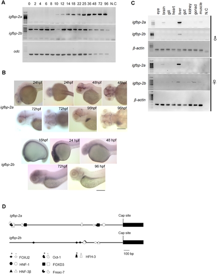
Temporal and spatial expression patterns of igfbp-2a and igfbp-2b. A) RT-PCR analysis result. The developmental stages are shown at the top, hpf, hour post fertilization. N.C., negative control. odc, (ornithine decarboxylase). B) In situ hybridization analysis of whole mounted embryos. Embryos of indicated stages were analyzed. Scale bar = 100 μm. C) Tissue distribution of igfbp-2a and igfbp-2b mRNA in male and female adult fish. D) Schematic diagram comparing the 5′-flanking region of igfbp-2a and igfbp-2b 2,000 bp before the cap site are shown. Close symbols indicate DNA binding elements in forward orientation and open symbols indicate those in reverse orientation. |
|
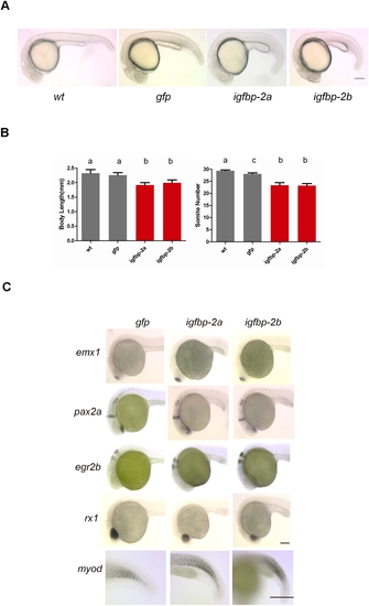
Ectopic expression of IGFBP-2a and IGFBP-2b causes a similar degree of reduction in embryonic growth and development but has no effect on cell fate and patterning. A) Representative images of wild-type, GFP mRNA (800 pg/embryo), IGFBP-2a mRNA (800 pg/embryo), or IGFBP-2b mRNA 800 pg/embryo) injected embryos at 24 hpf. B) Body length and somite number of the groups indicated. Results are from three independent microinjection experiments. The total embryo number for each group is 79 (wt), 95(gfp), 79(igfbp-2a), and 100 (igfbp-2b). Values are represented as means±S.E. (n = 3). Groups with common letters are not significantly different from each other (p<0.05). C) Whole mount in situ hybridization analysis of various marker genes in gfp mRNA-injected control (left column), igfbp-2a mRNA-injected (central column), and igfbp-2b mRNA-injected (right column) embryos at 24 hpf: emx1 expression in the forebrain; pax2a expression in the optic stalk, mid-hindbrain boundary, and hindbrain; egr2b expression in the third and fifth rhombomeres of the hindbrain; rx1 expression in the retina; myoD expression in the somatic myotome. Scale bar = 100 μm. Similar patterns were observed in all embryos examined in each group (n = 8–14). EXPRESSION / LABELING:
|


