
- Title
-
Zebrafish homologue irx1a is required for the differentiation of serotonergic neurons
- Authors
- Cheng, C.W., Yan, C.H., Choy, S.W., Hui, M.N., Hui, C.C., and Cheng, S.H.
- Source
- Full text @ Dev. Dyn.
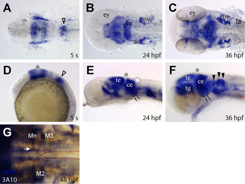
Developmental expression of irx1a in the zebrafish brain. A-F: Dorsal view of flat-mounted (A-C) and a lateral view (D-F) of the zebrafish brain region. A,D: irx1a is expressed in the prospective midbrain and the anterior hindbrain regions but not in the midbrain-hindbrain boundary at the five-somite stage (5 s). Note that irx1a is also weakly expressed in the posterior hindbrain region (open arrowheads). B,E: irx1a is expressed in the tectum, cerebellum, rostral hindbrain region, and acoustic ganglion at 24 hpf. C,F: By 36 hpf, expression of irx1a can be detected in the lateral line. In the hindbrain, irx1a expression is extended to the caudal part of hindbrain and in the commissural neurons (black arrowheads). G: Dorsal view of the hindbrain region of flat-mounted 48 hours postfertilization (hpf) embryo; double in situ hybridizaton and immunostaining staining of irx1a (dark blue) and 3A10 (brown). irx1a is not expressed in the reticulospinal neurons, and the Mauthner axons (white arrow) lie just dorsal to the ventral irx1a expression domain. Asterisks (*) indicate the position of midbrain-hindbrain boundary. ac, acoustic ganglion; ce, cerebellum; ey, eye; la, lateral line; Mn, Mauthner neuron; M2, Mi2cm; M3, Mi3cm; ov, otic vesicle; r1, rhombomere 1; te, tectum; tg, tegmentum. EXPRESSION / LABELING:
|
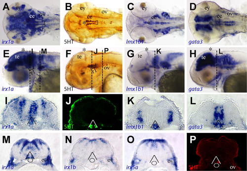
Expression of irx1a in hindbrain serotonergic neurons. A-H: Dorsal view of flat-mounted (A-D) and a lateral view (E-H) of a 48 hpf zebrafish brain region. A,B,E,F: irx1a is expressed in two rows of cells in rostral ventral hindbrain (black brackets, A,E) and have the same pattern as serotonergic neurons (B,F). C,D,G,H: The serotonergic markers Lmx1b1 (C,G) and Gata3 (D,H) are also expressed in the rostral ventral hindbrain. I-L: Transverse section of zebrafish hindbrain at rhombomere 1 position which are indicated in E-H, respectively. J: The serotonergic neurons are located in the ventral hindbrain (dashed lines) and irx1a is expressed in the same region. K: Lmx1b1 is expressed in the serotonergic neurons as well as in the floor plate. On the other hand, Gata3 is expressed in the serotonergic neurons but not the floorplate, and note that the ventral expression domain of Gata3 is boarder than the serotonergic neurons. M,P: Transverse section of zebrafish hindbrain at the otic vesicle region, which is indicated in E,F, respectively. The ventral expression domain of irx1a is on the boundary of notochord (dashed line circles in M) and is overlapped with serotonergic neurons (P). N,O: The other iro1 homologue irx1b (N) nor irx5a (O) are not expressed in this domain (N). EXPRESSION / LABELING:
|

Suppression of serotonergic (5HT) neuron formation by irx1a inactivation. A-D,I-K: Embryos are injected with control morpholino (MO). E-H,M-P: Embryos are injected with irx1a MO. A,E: Lateral view of 48 hours postfertilization (hpf) rostral hindbrain region. B,C,F,G: Dorsal view, dashed lines demarcate the eye, and asterisks (*) indicate the position of midbrain-hindbrain boundary. The differentiation of serotonergic neurons is examined by the expression of 5HT and tphR. The number of differentiated serotonergic neurons in the hindbrain is largely reduced in the irx1a morphant (E-G) in comparison to control MO-injected embryos (A-C). D,H: Acridine orange fluorescent staining (lateral view of 36 hpf embryos, anterior to the left) showed that irx1a morphant (H) has ectopic cell death at rostral ventral hindbrain. I-N: Dorsal view of hindbrain region. Hindbrain segmentation is visualized by znp-1 staining. I,L: The segmentation is not altered in irx1a morphant. J,K,M,N: Mauthner neurons in rhombomere 4 (J,M, black arrows indicate the crossing of Mauthner axons) and cranial facial motor neurons (K,N) are also differentiated and patterned normally in irx1a morphants, except a mild reduction of the motor neurons in some of the morphants. |
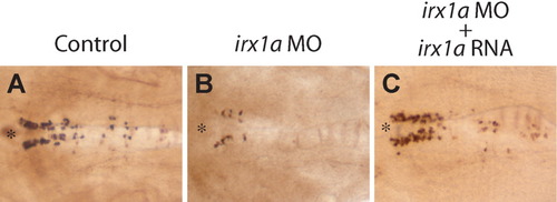
Figure 4. Rescue of irx1a morphant phenotypes by co-injection of irx1a mRNA. A-C: Dorsal view of a flat-mounted 48 hours postfertilization (hpf) zebrafish brain region. Embryos injected with irx1a morpholino (MO, B) show reduction of serotonergic neurons, whereas embryos co-injected with irx1a MO plus irx1a mRNA (C) show normal differentiation comparable to the control MO-injected embryos (A). |
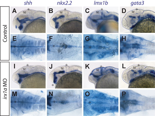
Early hindbrain development in the ventral domain is not affected in irx1a morphant. A-P: Lateral view (A-D,I-L) and dorsal view (E-H,M-P) of 30 hours postfertilization (hpf) zebrafish brain region. Control embryos (A-H) are not different from irx1a morphants (I-P) in floorplate expression of shh (A,E,I,M), ventral hindbrain expression of Nkx2.2 (B,F,J,N), Lmx1b1 (C,G,K,O), and Gata3 (D,H,L,P). Asterisks (*) indicate the position of the midbrain-hindbrain boundary. EXPRESSION / LABELING:
|