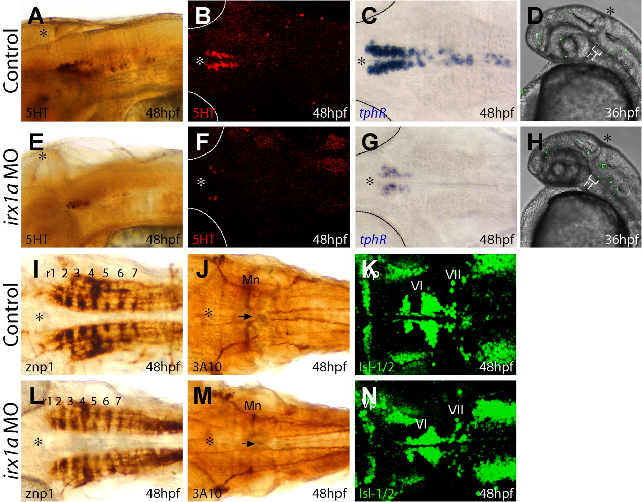
Fig. 3 Suppression of serotonergic (5HT) neuron formation by irx1a inactivation. A-D,I-K: Embryos are injected with control morpholino (MO). E-H,M-P: Embryos are injected with irx1a MO. A,E: Lateral view of 48 hours postfertilization (hpf) rostral hindbrain region. B,C,F,G: Dorsal view, dashed lines demarcate the eye, and asterisks (*) indicate the position of midbrain-hindbrain boundary. The differentiation of serotonergic neurons is examined by the expression of 5HT and tphR. The number of differentiated serotonergic neurons in the hindbrain is largely reduced in the irx1a morphant (E-G) in comparison to control MO-injected embryos (A-C). D,H: Acridine orange fluorescent staining (lateral view of 36 hpf embryos, anterior to the left) showed that irx1a morphant (H) has ectopic cell death at rostral ventral hindbrain. I-N: Dorsal view of hindbrain region. Hindbrain segmentation is visualized by znp-1 staining. I,L: The segmentation is not altered in irx1a morphant. J,K,M,N: Mauthner neurons in rhombomere 4 (J,M, black arrows indicate the crossing of Mauthner axons) and cranial facial motor neurons (K,N) are also differentiated and patterned normally in irx1a morphants, except a mild reduction of the motor neurons in some of the morphants.
Image
Figure Caption
Figure Data
Acknowledgments
This image is the copyrighted work of the attributed author or publisher, and
ZFIN has permission only to display this image to its users.
Additional permissions should be obtained from the applicable author or publisher of the image.
Full text @ Dev. Dyn.

