
Figure 5
- ID
- ZDB-FIG-260427-39
- Publication
- Choi et al., 2026 - 3D imaging with enhanced transparency, signal-to-background ratios, and antigen detection using HyPer-3D
- Other Figures
- All Figure Page
- Back to All Figure Page
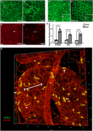
3D interrogation of poorly recognized antigens using HyPer-3D (A–F) Sections of the same testis were left as untreated controls (Cont) or treated with HyPer-3D (HyP), and then formamide/glycerol cleared after IF for the adult stem cell markers cKIT (A and B, green), STRA8 (C and D, green; inset shows high magnification, arrows highlight stained cells, and asterisks indicate regions of background noise), and GFR-α1 (E and F, red). Scale bars, 20 μm. (G) SBRs (∗∗∗ (H) 3D imaging of testis tubules for MCAM (red) and GFR-α1 (green) enabled visualization of spermatogonial stem cells in A-paired spermatogonia (arrows) and A-aligned spermatogonia at 4-cell (arrow heads) or 8-cell (bracket) stages of differentiation. |