
Figure 6
- ID
- ZDB-FIG-260427-41
- Publication
- Choi et al., 2026 - 3D imaging with enhanced transparency, signal-to-background ratios, and antigen detection using HyPer-3D
- Other Figures
- All Figure Page
- Back to All Figure Page
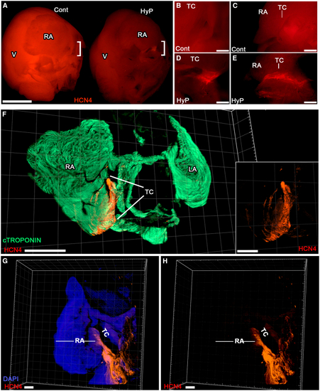
3D interrogation of muscular organs with enhanced clarity (A–E) IF performed on whole atria for HCN4 (red) that marks the sinoatrial node (SAN). Atria processed using standard H2O2 treatment (control, Cont) or HyPer-3D (HyP) were imaged side-by-side for comparison (A; scale bars, 2 mm, RA, right atria; LA, left atria). High-magnification analyses of bracketed regions in (A) showed a distinct HCN4+ node at the terminal crest (TC) of hearts imaged with HyPer-3D (D, coronal view; E, lateral view), which was not readily discernible in control hearts (B, coronal view; C, lateral view). Scale bars, 0.25 mm. (F) 3D confocal microscopy imaging of formamide/glycerol-cleared whole atria immunolabeled for both the working myocardium (cTROPONIN-T, green) and HCN4 pacemaker channels (red; scale bars, 1 mm). Inset shows SAN alone. (G and H) Staining with DAPI and HCN4 (G, merged; H, HCN4 alone; scale bars, 0.2 mm). |