|
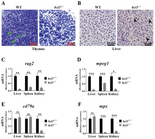
Histological abnormalities and reduced immune cell marker expression in bcl3−/− zebrafish. (A) H&E-stained sections of thymus from WT and bcl3−/− zebrafish at 4 months of age. Green arrows indicate Hassall bodies. (B) H&E-stained sections of liver from WT and bcl3−/− zebrafish. Black arrows indicate hepatocytes with nuclear displacement and pyknosis. Relative mRNA expression of (C) rag2 (T cell marker), (D) mpeg1 (macrophage marker), (E) cd79a (B cell marker), and (F) mpx (neutrophil marker) in the liver, spleen, and kidney of bcl3+/+ and bcl3−/− zebrafish at 4 months of age. *** p < 0.001, ** p < 0.01, * p < 0.05 (t-test), ns indicates not significant. Error bars represent SD.
|