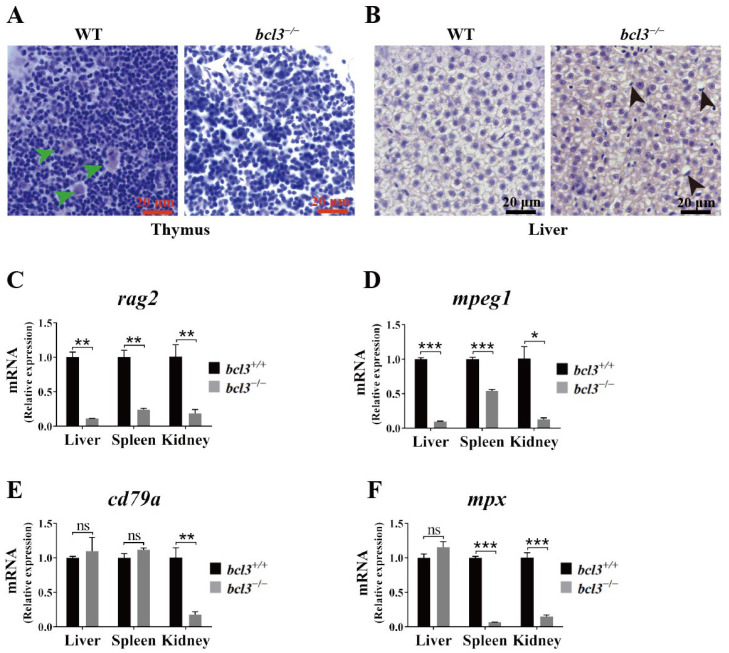
IMAGE

Figure 3

ID
ZDB-IMAGE-251226-9
Genes
Source
Figures for Fan et al., 2025
Image
Figure Caption

Figure 3

Histological abnormalities and reduced immune cell marker expression in bcl3−/− zebrafish. (A) H&E-stained sections of thymus from WT and bcl3−/ zebrafish at 4 months of age. Green arrows indicate Hassall bodies. (B) H&E-stained sections of liver from WT and bcl3−/ zebrafish. Black arrows indicate hepatocytes with nuclear displacement and pyknosis. Relative mRNA expression of (C) rag2 (T cell marker), (D) mpeg1 (macrophage marker), (E) cd79a (B cell marker), and (F) mpx (neutrophil marker) in the liver, spleen, and kidney of bcl3+/+ and bcl3−/− zebrafish at 4 months of age. *** p < 0.001, ** p < 0.01, * p < 0.05 (t-test), ns indicates not significant. Error bars represent SD.

Figure Data
Acknowledgments
This image is the copyrighted work of the attributed author or publisher, and ZFIN has permission only to display this image to its users. Additional permissions should be obtained from the applicable author or publisher of the image. Full text @ Cells