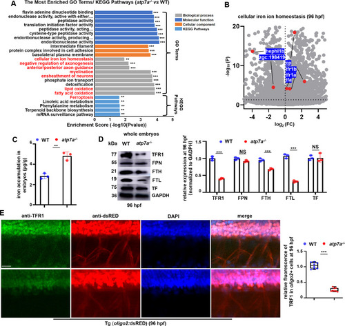
Atp7a deficiency induces significant changes in iron homeostasis. (A) Go terms and KEGG pathways for the differentially expressed genes (DEGs) in atp7a−/− larvae at 96 hpf, such as cellular iron ion homeostasis, myelination, ferroptosis, etc., were enriched. (B) Scatter plots for cellular iron ion homeostasis genes in atp7a−/− larvae at 96 hpf. (C) The iron content (μg/g) in WT and atp7a−/− at 96 hpf. Each dot represents one repeat. (D) Western blotting (WB) analysis of the relative protein level of TFR1, FPN, FTH, FTL, and TF in WT or atp7a−/− whole larvae at 96 hpf, with GADPH as an internal control (graphs). Each dot represents one repeat. The calculation assay is followed for the next western blotting data. (E) Double staining of oligo2+ cells (RED) with TFR1 (GFP) in WT or atp7a−/− larvae at 96 hpf, and the calculation of the relative TFR1 fluorescence intensity in oligo2+ cells (box and whisker plots). Each dot represents the relative level of signal in a representative image in an individual embryo or larvae in each group. The calculation assay is followed for the next immunofluorescence data. Scale bar: 50 μm (E). *P < 0.05, **P < 0.01, ***P < 0.001. NS, not significant. (For interpretation of the references to colour in this figure legend, the reader is referred to the web version of this article.)
|
