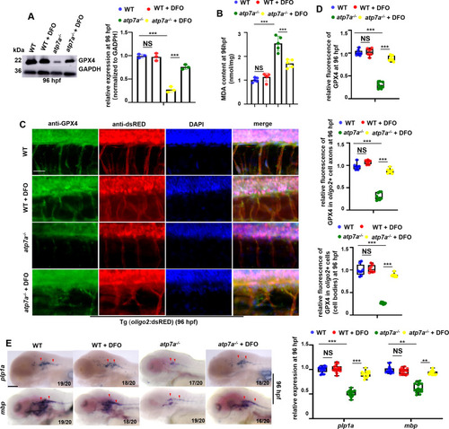
Iron chelator DFO can effectively restore GPX4 expression and myelin and axon developmental defects in atp7a−/−. (A) The relative protein level of GPX4 in WT and atp7a−/− larvae treated with or without DFO at 96 hpf, with GADPH as an internal control (graphs). (B) MDA content in WT and atp7a−/− larvae treated with or without DFO at 96 hpf. (C) Double staining of oligo2+ cells (RED) with GPX4 (GFP) in WT or atp7a−/− larvae from groups with or without DFO treatment at 96 hpf. (D) The calculation of the relative fluorescence level of GPX4 (up), GPX4 fluorescence intensity in oligo2+ cell axons (middle), and GPX4 fluorescence intensity in oligo2+ cell bodies (down) (box and whisker plots). (E) Transcriptional expressions of CNS myelin genes mbp and plp1a in WT or atp7a−/− larvae from groups with or without DFO treatment at 96 hpf, and the calculation of the relative expressions of mbp and plp1a (box and whisker plots) at 96 hpf. E, lateral view, anterior to the left. Scale bar: 50 μm (C), 100 μm (E). *P < 0.05, **P < 0.01, ***P < 0.001. NS, not significant. (For interpretation of the references to colour in this figure legend, the reader is referred to the web version of this article.)
|
