
Fig. 2
- ID
- ZDB-FIG-250818-31
- Publication
- Kawasaki et al., 2025 - Meioc-Piwil1 complexes regulate rRNA transcription for differentiation of spermatogonial stem cells
- Other Figures
- All Figure Page
- Back to All Figure Page
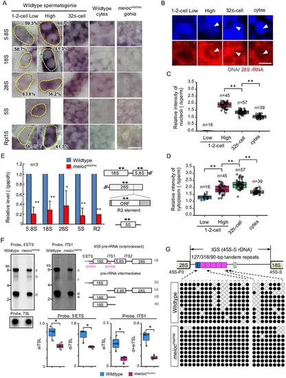
Defect on upregulation of rRNA transcription in meiocmo/mo spermatogonia. (A) In situ hybridization of 5S, 5.8S, 18S, and 28S rRNA and immunohistochemistry with anti-Rpl15 antibody in spermatogonia (gonia) and spermatocytes (cyte) in wild-type and meiocmo/mo. Yellow dotted lines indicate 1- to 2-cell spermatogonia. Percentages represent the frequency of low and high 1- to 2-cell spermatogonia. (B–D) Fluorescent in situ hybridization of 28S rRNA in wild-type. The graphs present quantification of signal intensities of 28S rRNA in nucleoli (C) and in cytoplasm (D) in spermatogenic cells. Arrowheads: nucleoli; cytes: spermatocytes. (E) qRT-PCR analysis of rRNAs and R2 between wild-type and meiocmo/mo purified sox17::egfp positive spermatogonia. Two-way arrows in the schema indicate the position of primers on the rRNA and R2 element. (F) Northern blot analysis of pre-rRNA processing in wild-type and meiocmo/mo testes using probes for 5’ external transcribed spacer (ETS) and internal transcribed spacer 1 (ITS1). Right panel: schema of 45S pre-rRNA and pre-rRNA processing intermediates in zebrafish (Tao et al., 2017). Left panel: Northern blot analysis of pre-rRNA processing in wild-type and meiocmo/mo testes using 5’ETS and ITS1 probes. A probe for the 7SL RNA was used as a loading control. Graphs summarize relative signal intensity of 45S pre-rRNA and intermediates normalized to 7SL in three wild-type and meiocmo/mo testes. (G) Bisulfite-sequencing analysis of the tandem repeat region in the intergenic spacer (IGS) region of the 45S-S rDNA locus in purified undifferentiated spermatogonia of wild-type and meiocmo/mo. Arrows: position of bisulfite primers in the tandem repeat elements (blue, magenta, and white boxes); black dots: methylated CpG sites; white dots: unmethylated sites. *p<0.05, **p<0.01. Scale bars: 10 µm. |