
Fig. 3 - Supplemental 1
- ID
- ZDB-FIG-250818-35
- Publication
- Kawasaki et al., 2025 - Meioc-Piwil1 complexes regulate rRNA transcription for differentiation of spermatogonial stem cells
- Other Figures
- All Figure Page
- Back to All Figure Page
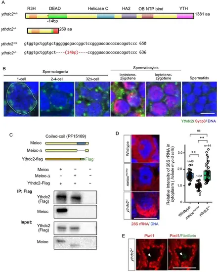
Mutations of the ythdc2-/-. (A) Zebrafish Ythdc2 protein structure and mutation sequence (*) in the ythdc2 KO zebrafish. (B) Double staining of a testis with anti-Ythdc2 antibody (green) and anti-Sycp3 antibody (red). Staging of spermatogonia was defined by the number of cells in the cyst, and staging of spermatocytes was defined by the patterns of Sycp3. Scale bar: 5 µm. (C) Pull-down assay using full-length or coiled-coil domain-deleted Meioc and Flag-tagged Ythdc2. Schema: conditions of coexpression of each protein in HEK-293A cells used for the pull-down assay. (D) In situ hybridization of 28S rRNA (left panels) and quantification of the 28S rRNA signal intensities (right panels) in wild-type, meiocmo/mo, and ythdc2-/- 1- to 2-cell spermatogonia. The cytoplasmic signal intensities were normalized to the myoid cell cytoplasm. **p<0.01, ns: not significant. (E) Immunostaining against Piwil1 (red) and fibrillarin (green) in ythdc2-/- spermatogonia. Arrowheads: undetectable Piwil1 signals in fibrillarin (green) positive nucleoli under the normal sensitivity imaging of Piwil1. |