
Fig. 3 - Supplemental 4
- ID
- ZDB-FIG-250808-51
- Publication
- Wu et al., 2025 - Pu.1/Spi1 dosage controls the turnover and maintenance of microglia in zebrafish and mammals
- Other Figures
-
- Fig. 1
- Fig. 2
- Fig. 2 - Supplemental 1
- Fig. 3
- Fig. 3 - Supplemental 1
- Fig. 3 - Supplemental 2
- Fig. 3 - Supplemental 3
- Fig. 3 - Supplemental 4
- Fig. 3 - Supplemental 5
- Fig. 3 - Supplemental 6
- Fig. 4
- Fig. 5
- Fig. 5 - Supplemental 1
- Fig. 5 - Supplemental 2
- Fig. 5 - Supplemental 3
- Fig. 6
- Fig. 6 - Supplemental 1
- All Figure Page
- Back to All Figure Page
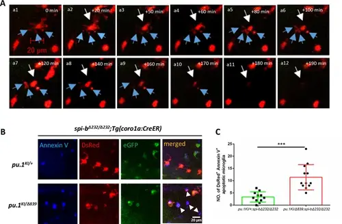
Pu.1/Spi-b-deficient microglia undergo apoptosis in zebrafish. (A) Time-lapse live imaging shows the blebbing and fragmentation of DsRed+ microglia between 3 dpf and 5 dpf in pu.1KI/Δ839;spi-bΔ232/Δ232;Tg(coro1a:CreER) embryos treated with 4-OHT from 48 hpf to 60 hpf. The blue arrows indicate the formation of apoptotic cell bodies. (B) Fluorescent live imaging of Annexin V, DsRed and eGFP signals in 4-dpf pu.1KI/+;spi-bΔ232/Δ232;Tg(coro1a:CreER) and pu.1KI/Δ839;spi-bΔ232/Δ232;Tg(coro1a:CreER) embryos treated with 4-OHT from 48 hpf to 60 hpf and subjected to brain injection of Annexin V-647. (C) Quantification of DsRed+Annexin+ microglia in (B). (pu.1KI/+;spi-bΔ232/Δ232;Tg(coro1a:CreER) n=11, pu.1KI/Δ839;spi-bΔ232/Δ232;Tg(coro1a:CreER) n=12) ***p<0.001. |