
FIGURE
Fig. 3 - Supplemental 2
- ID
- ZDB-FIG-250808-49
- Publication
- Wu et al., 2025 - Pu.1/Spi1 dosage controls the turnover and maintenance of microglia in zebrafish and mammals
- Other Figures
-
- Fig. 1
- Fig. 2
- Fig. 2 - Supplemental 1
- Fig. 3
- Fig. 3 - Supplemental 1
- Fig. 3 - Supplemental 2
- Fig. 3 - Supplemental 3
- Fig. 3 - Supplemental 4
- Fig. 3 - Supplemental 5
- Fig. 3 - Supplemental 6
- Fig. 4
- Fig. 5
- Fig. 5 - Supplemental 1
- Fig. 5 - Supplemental 2
- Fig. 5 - Supplemental 3
- Fig. 6
- Fig. 6 - Supplemental 1
- All Figure Page
- Back to All Figure Page
Fig. 3 - Supplemental 2
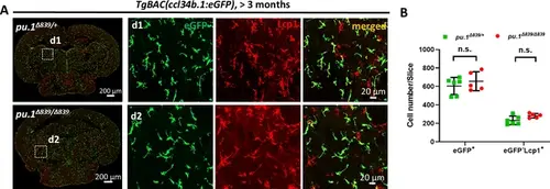
Microglia number is not affected in pu.1Δ839 null mutants. (A) Representative images show the co-staining of eGFP and Lcp1 antibodies on the midbrain cross section of adult pu.1Δ839/+;TgBAC(ccl34b.1:eGFP) and pu.1Δ839/Δ839;TgBAC(ccl34b.1:eGFP) fish. eGFP+ cells represent microglia, whereas eGFP-Lcp1+ cells are dendritic cells (DCs). (B) Quantification of the number of eGFP+ microglia and eGFP-Lcp1+ DCs on the midbrain cross section of adult pu.1Δ839/+;TgBAC(ccl34b.1:eGFP) (n=6) and pu.1Δ839/Δ839;TgBAC(ccl34b.1:eGFP) (n=5) fish. n.s.=not significant, p>0.05. |
Expression Data
Expression Detail
Antibody Labeling
Phenotype Data
Phenotype Detail
Acknowledgments
This image is the copyrighted work of the attributed author or publisher, and
ZFIN has permission only to display this image to its users.
Additional permissions should be obtained from the applicable author or publisher of the image.
Full text @ Elife