|
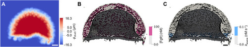
Spatially varying sources and sinks in the 3D model at an exemplary time point at ≈60% epiboly. (A) Signed-distance function (SDF) of the embryo shell boundary, φshell, for an exemplary z-slice. Combining φshell (representing YSL and EVL boundaries) with φECS enables the restriction of sources and sinks to the deep-cell surfaces. (B,C) A clipped model of the ECS showing that Fgf receptors (B, pink) are restricted to the deep cell boundaries, i.e. not the EVL or YSL. The sources (C, blue) are restricted to ≈5 rows of deep cells above the margin of the blastoderm. Receptor concentrations are given in units of nM; ksource is given in nM/s. Scale bars: 50 μm.
|