FIGURE

Figure 7

ID
ZDB-FIG-250423-70
Publication
Reed et al., 2025 - Oxygen chemoreceptor inhibition by dopamine D2 receptors in isolated zebrafish gills
Other Figures
All Figure Page
Back to All Figure Page
Figure 7

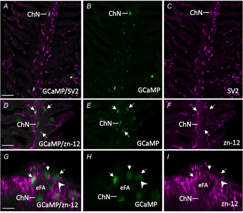
Characterization of GCaMP‐positive postsynaptic chain neurons (ChNs) in Tg(elavl3:GCaMP6s) zebrafish gills

AC, confocal imaging of immunohistochemical localization of GCaMP in a ChN containing synaptic vesicle protein‐2 (SV2, magenta). B and C, GCaMP and SV2 labelling shown separately. Scale bar = 50 µm in (A) to (C). DF, co‐labelling of GCaMP‐positive ChNs and NECs (green, arrows) with nerve fibres labelled with zn‐12 (magenta). E and F, GCaMP and zn‐12 labelling shown separately. Scale bar = 50 µm in (D) to (F). GI, images from (D) to (F), cropped and titled back 90°. Rotation demonstrates neural connection (arrowhead) between nerve fibres located below the efferent filament artery (eFA) where ChNs are located, projecting to GCaMP‐positive NECs. [Colour figure can be viewed at wileyonlinelibrary.com]

Expression Data

Expression Detail
Antibody Labeling
Phenotype Data

Phenotype Detail
Acknowledgments
This image is the copyrighted work of the attributed author or publisher, and ZFIN has permission only to display this image to its users. Additional permissions should be obtained from the applicable author or publisher of the image. Full text @ J. Physiol.