|
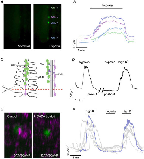
The chain neuron (ChN) calcium response to hypoxia requires synaptic contact with neuroepithelial cells (NECs) A, examples from live fluorescence imaging at 488 nm of four ChNs along a single filament in normoxia (left micrograph) and hypoxia (right micrograph). B, calcium traces from the four ChNs shown in (A) responding to hypoxia. Different colours represent corresponding cells (ChN 1–4) in (A). C, schematic of a gill filament (left) and rotation by 90° on the y‐axis (right) illustrate filament transection. Filaments were cut along the red dashed line at the proximal end where a ChN was present, but no NECs were observable. D, calcium imaging trace of a single ChN before and after filament transection. After the filament was cut (break in trace), the neuron no longer responded to hypoxia (n = 5 cells). As a positive control, viability of the neuron was demonstrated by stimulation with a solution of high extracellular K+. E, confocal imaging of gills from a double transgenic animal produced by crossing Tg(elavl3:GCaMP6s) and Tg(dat:tom20 MLS‐mCherry) fish showing the relationship between the dopamine active transporter (DAT) nerve endings (magenta) and GCaMP‐positive NECs (green) in control (left micrograph) and 6‐OHDA‐treated gills (right micrograph). DAT labelling was found in close proximity to NECs but was reduced after 6‐OHDA treatment. F, overlayed Ca2+ imaging traces from 6‐OHDA treated animals. After 6‐OHDA treatment, the ChNs did not respond to hypoxia (n = 6 cells). Response to high extracellular K+ confirmed cell viability. The schematic in (C) was created with BioRender.com. [Colour figure can be viewed at wileyonlinelibrary.com]
|