FIGURE

Figure 6.

ID
ZDB-FIG-250415-82
Publication
Torcq et al., 2025 - Single Molecule Fluorescence In Situ Hybridization Using RNAscope to Study Hematopoietic and Vascular Interactions in the Zebrafish Embryo and Larva
Other Figures
All Figure Page
Back to All Figure Page
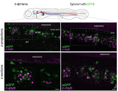
Figure 6.

Whole-mount in situ analysis of cmyb mRNA colocalization with HSPCs at 5 dpf.

Spinning disk confocal images obtained with a 5 dpf larva [Tg(runx1+23:eGFP) fish line] expressing eGFP in HSPCs under the control of the runx1+23 enhancer. Panels are constituted from images obtained either in the trunk region (left: AGM) or in the tail (right: CHT), either from maximum z-projections of z-stacks or from single z-sections with 2× magnification of regions in white boxes, highlighting hematopoietic clusters (dashed lines). RNAscope signals (magenta) colocalize in the majority with HSPCs (see also Figure 7 and Video 3 for 3D reconstitution). AGM: aorta gonad mesonephros; CHT: caudal hematopoietic tissue; HSPCs: hematopoietic stem and progenitor cells. Scale bars = 20 μm.

Expression Data

Expression Detail
Antibody Labeling
Phenotype Data

Phenotype Detail
Acknowledgments
This image is the copyrighted work of the attributed author or publisher, and ZFIN has permission only to display this image to its users. Additional permissions should be obtained from the applicable author or publisher of the image. Full text @ Bio Protoc