|
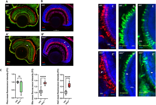
Sacsin deficiency alters cell fate and photoreceptor structure in zebrafish retina (A) Representative confocal images of the retina of sacs-/- zebrafish obtained after immunostaining of rods with a rhodopsin antibody (red) and with PKC-α to label bipolar cells. (B) Immunostaining of amacrine cells with a HuC antibody (red). Nuclei were highlighted with DAPI·(C) Analysis of fluorescence mean intensity in knockout larvae compared to controls. (D-F′) Representative confocal images of the retina of sacs-/- zebrafish obtained after immunostaining of rods with a rhodopsin antibody (red) and cones (Uv-opn1sw and Blue-opn1sw) in green. Scale bar = 50 μm (A-B′) and 10 μm (C-D′). (For interpretation of the references to colour in this figure legend, the reader is referred to the web version of this article.)
|