FIGURE

Fig. 4

ID
ZDB-FIG-250203-33
Publication
Naef et al., 2025 - Modeling sacsin depletion in Danio Rerio offers new insight on retinal defects in ARSACS
Other Figures
All Figure Page
Back to All Figure Page
Fig. 4

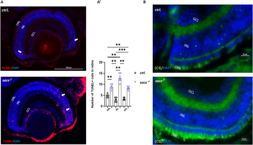
Increased apoptosis contributes to retinal defects in sacs-/- zebrafish mutants. (A) Representative TUNEL staining on retinal frozen section of zebrafish larvae at 120 hpf from control embryos and mutants. (A') Quantification of the number of TUNEL-positive cells in the entire retina based on three independent measurements of larvae per group at 120 hpf. ** p < 0.01; *** p < 0.001 were calculated by Kruskal Wallis test multiple comparisons. (B) Immunostaining of the retinal section with activated Caspase-3 antibody (green) at 120 hpf. Nuclei were highlighted with DAPI (blue). ONL, outer nuclear layer; INL, inner nuclear layer, GCL, ganglion cell layer. Scale bar = 100 μm (A) and 10 μm (B). All images were acquired using a Zeiss LSM 900 confocal-microscope. TUNEL+ cells were counted per area considering whole retina section. (For interpretation of the references to colour in this figure legend, the reader is referred to the web version of this article.)

Expression Data

Expression Detail
Antibody Labeling
Phenotype Data
Fish:
Observed In:
Stage: Day 5

Phenotype Detail
Acknowledgments
This image is the copyrighted work of the attributed author or publisher, and ZFIN has permission only to display this image to its users. Additional permissions should be obtained from the applicable author or publisher of the image. Full text @ Neurobiol. Dis.