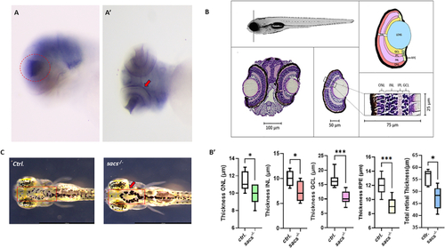
Loss of sacs gene affect neural retina formation and altered VBA reflex. (A) At 72 hpf sacs gene is expressed in the most anterior region of the CNS and in the eye field (red circle in A, lateral view; red arrow in A', ventral view). (B-B′) Hematoxylin/eosin staining revealed a normal organization in retina structure in sacs-/- larvae and reduced thickness of ONL (the outer nuclear layer), INL (inner nuclear layer), GCL (ganglion cell layer), and in RPE (retinal pigmented epithelium) and in total retinal thickness. Error bars indicate mean ± s.e.m. Statistical differences were computed using the two-tailed Mann-Whitney test and are indicated as (*p < 0.05; ***p < 0.001). (IPL (inner plexiform layer)). Results were obtained from representative sections from 5 zebrafish control embryos and 5 sacs-/- mutant larvae. C) Head dorsal view of 120 hpf control and sacs-/- larvae with insets showing their pigmentation pattern (red arrow). Scale bar = 100 μm. (For interpretation of the references to colour in this figure legend, the reader is referred to the web version of this article.)
|
