
FIGURE
Figure 7
Figure 7
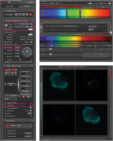
Confocal microscopy setting The left panel shows the overview of standard setting for confocal microscopy. The settings in pink boxes are essential to be able to generate images that can be used for quantification with Cellpose. This includes an image format of 1024 × 1024 with a pixel size of 0.38 μm. The upper right panel shows laser settings to detect cells. The lower right panel shows an overview of example zebrafish brain with zebrafish nuclei in cyan and human cells in yellow. |
Expression Data
Expression Detail
Antibody Labeling
Phenotype Data
Phenotype Detail
Acknowledgments
This image is the copyrighted work of the attributed author or publisher, and
ZFIN has permission only to display this image to its users.
Additional permissions should be obtained from the applicable author or publisher of the image.
Full text @ STAR Protoc