
Figure 5
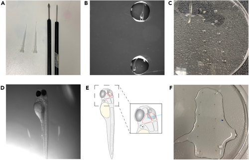
Prepare and mount larvae for injection (A) Shows utensils used for positioning and later removal of agarose. (B) Shows larvae placed in a drop of medium in the lid of a 10 cm Petri dish. (C) Shows an overview of larvae in drops of medium. Larvae are lined up from top to bottom and in rows to later facilitate the injection procedure. (D) Shows a larva positioned in agarose. Note the slight tilt of the body to the right lateral side. (E) Shows a schematic of a positioned larva with the midbrain outlined in red as well as the brain enlarged to show correct needle positioning. (F) Shows agarose-embedded larvae with added E3 and 1x tricaine solution on top. |