FIGURE

Figure 4

ID
ZDB-FIG-241219-146
Publication
Testa et al., 2024 - Hypoxic Human Microglia Promote Angiogenesis Through Extracellular Vesicle Release
Other Figures
All Figure Page
Back to All Figure Page
Figure 4

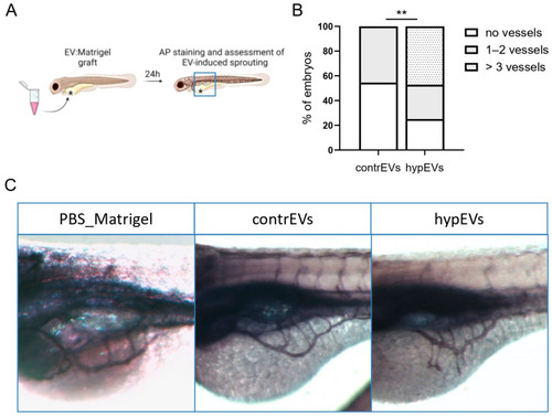
Analysis of the angiogenic potential of microglial EVs in zebrafish. (A) Schematic of the experimental set-up. EVs were grafted with Matrigel in the proximity of the developing sub-intestinal vein (SIV), as shown by a black asterisk, and vascular sprouting from the SIV was assessed after 24 h. The area of vessel analysis is represented by a blue square. (B) Histograms represent the number of vessels sprouting towards the implant, counted for each embryo, divided into categories (no vessels, 1–2 vessels, >3 vessels). Data are expressed as the percentage of embryos in each category (n = 36 embryos/group). (C) Representative confocal images showing whole-mount alkaline phosphatase (AP) staining of embryos at 1 dpi in lateral view. Injection of hypEVs stimulated the migration of SIV-derived vascular sprouts towards the implant, while injection with contrEVs resulted in a normal development of the vascular plexus. Chi-Square test was performed. ** p < 0.01.

Expression Data

Expression Detail
Antibody Labeling
Phenotype Data

Phenotype Detail
Acknowledgments
This image is the copyrighted work of the attributed author or publisher, and ZFIN has permission only to display this image to its users. Additional permissions should be obtained from the applicable author or publisher of the image. Full text @ Int. J. Mol. Sci.