FIGURE

Figure 7

ID
ZDB-FIG-241219-149
Publication
Testa et al., 2024 - Hypoxic Human Microglia Promote Angiogenesis Through Extracellular Vesicle Release
Other Figures
All Figure Page
Back to All Figure Page
Figure 7

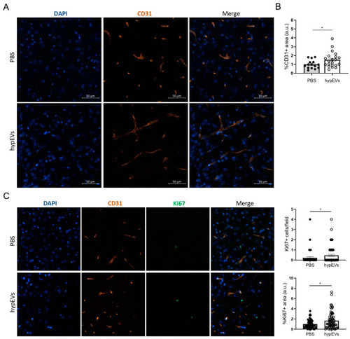
Evaluation of the hypEV effects on brain angiogenesis in a mouse model for ischemic stroke. After MCAO, mice were injected intraperitoneally with PBS or hypEVs, immediately after the procedure and each 24 h, for a total of 5 consecutive days. Mice were perfused at day 7 post-MCAO (n = 5 PBS, n = 6 hypEVs). (A) Representative pictures of blood vessels in the perilesional area. Immunofluorescence on brain sections was performed using anti-CD31 antibodies (endothelial cells) and Hoechst (nuclei). Images were acquired at a confocal microscope, using a 25× objective. Scale bar: 50 µm. (B) Quantification of the CD31 signal in the perilesional region was performed using ImageJ, fixing a threshold to detect the percentage of the CD31+ area, expressed as arbitrary units (a.u.), for each field of view analysed. (C) Analysis of proliferating nuclei in the perilesional area. The number of proliferating cells per field of view analysed is reported. Quantification of the Ki67+ area was performed using ImageJ and is expressed as normalised to controls. Bars represent mean ± SEM. The non-parametric t-test was applied. * p < 0.05.

Expression Data

Expression Detail
Antibody Labeling
Phenotype Data

Phenotype Detail
Acknowledgments
This image is the copyrighted work of the attributed author or publisher, and ZFIN has permission only to display this image to its users. Additional permissions should be obtained from the applicable author or publisher of the image. Full text @ Int. J. Mol. Sci.