
Fig. EV4
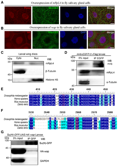
mRpL4 and wap may function through Su(H) to regulate Notch signaling A, B. Representative images of third instar larvae salivary glands (n > 10 larvae) from flies expressing FLAG‐tagged mRpL4 (A) and HA‐tagged wap (B). Immunostaining was performed using anti‐FLAG and anti‐HA antibodies to reveal mRpL4 and wap, respectively. Mitochondria are marked by GFP and cell nuclei are labeled by DAPI. C. Representative western blotting (n = 3 biological replicates) of mRpL4 protein distribution in cytoplasmic (Cyto) and nuclear (Nuc) fractions from wing disks lysates. D. Representative immunoprecipitation analysis (n = 3 biological replicates) using lysates from wing disks expressing GFP‐tagged mnb. Anti‐GFP antibodies were used for immunoprecipitation. Western blotting was performed using anti‐GFP and anti‐mRpL4 antibodies to reveal mnb and mRpL4, respectively. α‐Tubulin was used as control. E. Alignment of Su(H) protein sequences from fly, human, mice and zebrafish. The region covering the mnb phosphorylation consensus sequence is shown, and the conserved Thr residue (T426) is labeled by red box. F. Alignment of Notch protein sequences from fly, human, mice and zebrafish. The region covering the mnb phosphorylation consensus sequence is shown, and the conserved Ser residue (S2659) is labeled by red box. G. Representative immunoprecipitation analysis (n = 2 biological replicates) using lysates from wing disks expressing GFP‐tagged Su(H) and HA‐tagged wap. Anti‐GFP antibodies were used for immunoprecipitation. Western blotting was performed using anti‐GFP and anti‐HA antibodies to reveal Su(H) and wap, respectively. GAPDH was used as control. |