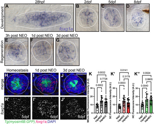
RNA in situ hybridization shows foxg1a expression in developing and regenerating posterior lateral line tissue. (A-D) Wholemount RNA in situ hybridization of foxg1a in wild-type zebrafish posterior lateral line primordium at 28 hpf (A), and in neuromasts at 2 dpf (B), 5 dpf (C), and 8 dpf (D). (F-G) Wholemount RNA in situ hybridization of foxg1a in wild-type zebrafish neuromasts during regeneration following neomycin (NEO) exposure at 5 dpf. (E) 3 h post-NEO, (F) 1-day post-NEO, and (G) 3 days post-NEO. (H-J′) Confocal projections of wild-type neuromasts showing hair cells labeled with Tg(myo6:GFP) (green), foxg1a expression with HCR fluorescent in situ hybridization (red) and nuclei labeled with DAPI (blue) at 5 dpf (H), 1 day-post NEO exposure (I), and 3 days-post NEO (J). Quantification of HCR foxg1a fluorescence intensity in arbitrary units (A.U.) in the whole neuromasts (K), hair cells (hc; K′), and support cells (sc; K″). n=15 NMs (nine larvae) 5 dpf, n=16 NMs (nine larvae) 1 day-post NEO, and n=18 NMs (nine larvae) 3 days-post NEO). All data presented at mean±s.d. Kruskal–Wallis test with Dunn's multiple comparisons. Scale bars: 20 µm.
|
