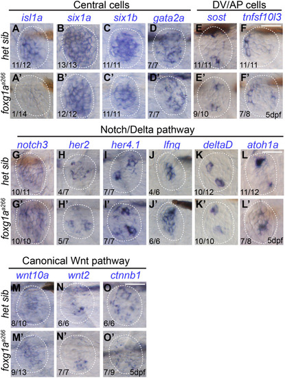
RNA in situ hybridization of NM cell markers. DIC images of wholemount RNA in situ hybridization showing mRNA expression in NMs at 5 dpf in heterozygous sibling (het sib) and foxg1aa266 larvae, lower lefthand numbers indicate the number of larvae with expression in neuromasts over the total number analyzed. Central cell markers: isl1a expression in 11/12 het sib (A) and 1/14 foxg1aa266 larvae (A′), six1a expression in 13/13 het sib (B) and 12/12 foxg1aa266 larvae (B′), six1b expression in 11/11 het sib (C) and 11/11 foxg1aa266 larvae (C′), and gata2a expression in 7/7 het sib (D) and 7/7 foxg1aa266 larvae (D′). Dorsoventral and anterior-posterior (DV/AP) cell markers: sost expression in 11/11 het sib (E) and 9/10 foxg1aa266 larvae (E′) and tnfs10l3 expression in 11/11 het sib (F) and 7/8 fox1gaa266 larvae (F′). Expression of Notch/Delta pathways markers: notch3 expression in 10/11 het sib (G) and 10/10 foxg1aa266 larvae (G′), her7 expression in 4/7 het sib (H) and 5/7 foxg1aa266 larvae (H′), her4.1 expression in 7/7 het sib (I) and 7/7 foxg1aa266 larvae (I′), lfng expression in 4/6 het sib (J) and 6/6 foxg1aa266 larvae (J′), deltaD expression in 10/12 het sib (K) and 10/10 foxg1aa266 larvae (K′), and atoh1a expression in 11/12 het sib (L) and 7/8 foxg1aa266 larvae (L′). Canonical Wnt pathway: wnt10a expression in 8/10 het sib (M) and 9/13 foxg1aa266 larvae (M′), wnt2 expression in 6/6 het sib (N) and 7/7 foxg1aa266 larvae (N′), and ctnnb1 expression in 6/6 het sib (O) and 7/9 foxg1aa266 larvae (O′). Scale bars: 20 µm.
|
