|
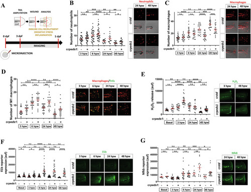
Peds1 deficiency impairs inflammation resolution and causes aberrant immune cells recruitment in a sterile tail injury model. A Schematic of the experimental procedure used for tail injury assays. One-cell stage zebrafish eggs were microinjected with std or peds1 crRNA/Cas9 complexes. At 3 dpf larvae were tail amputated and then inflammation and immune cell recruitment at the injury site (dotted lines) were analyzed at 3, 6, 24 and 48 h post-wounding (hpw) by fluorescence microscopy. The wound area is defined as the region between the amputation edge and the end of the caudal hematopoietic tissue (CHT). B, C Number of neutrophil and macrophages recruited at the injury site and representative merge images (brightfield and red channel; Tg(lyz:DsRED2)nz50 and Tg(mfap4.1:Tomato)xt12, respectively). D Number of M1-like macrophages (Tnfa+) recruited at the injury site and representative merge images (red channel: Tg(mfap4.1:Tomato)xt12 and green channel: Tg(tnfa:eGFP-F)ump5). E Analysis of H2O2 release at the wound site using the fluorogenic substrate acetyl-pentafluor-obenzene sulphonyl fluorescein. E–G Analysis and representative images of Il1b (green channel; Tg(il1b:GFP-F)ump3) and Nfkb fluorescent reporter expression levels (green channel; Tg(NFkB-RE:eGFP)sh235) at the wound site. Each point represents one larva and the mean ± SEM of each group is also shown. P-values were calculated using one-way ANOVA and Tukey’s multiple range. n.s, not significant, *p ≤ 0.05, **p ≤ 0.01, ***p ≤ 0.001, ****p ≤ 0.001. auf arbitrary units of fluorescence.
|