|
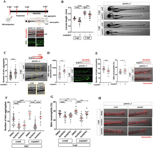
Peds1 deficiency exacerbates chronic skin inflammation. A Schematic of the experimental procedure used for chronic skin inflammation assays. Spint1a-deficient one-cell stage eggs were microinjected with std or peds1 crRNA/Cas9 complexes and imaged at 3 and 5 dpf. Treatments of interest were added to dechorionated embryos at 1 dpf by bath immersion and renewed daily. Spint1a-deficient larvae showed chronic skin inflammation with increased skin neutrophil infiltration, Nfkb fluorescent reporter expression levels and keratinocyte aggregates. B Analysis and representative images (brightfield channel) of larval length at 3 and 5 dpf. C Number of keratinocyte aggregates in the skin (red arrows) and representative images (brightfield channel) at 3 dpf. D Analysis and representative images of Nfkb fluorescent reporter expression levels (green channel; Tg(NFkB-RE:eGFP)sh235) at 3 dpf by fluorescence microscopy. E Total number and percentage of neutrophils in the skin and representative merge images (brightfield and red channel; Tg(lyz:DsRED2)nz50) quantitated by fluorescence microscopy. F Number of skin aggregates and (G) percentage of skin neutrophils quantitated in crstd and crpeds1 spint1a-/- larvae treated for 48 h with 20 µM HsVEPE1, 20 µM HsAEPE1 or vehicle (DMSO). H Representative merge images (brightfield and red channel; Tg(lyz:DsRED2)nz50) of each treatment analyzed in (F) and (G) by microscopy fluorescence are shown. Each point represents one larva and the mean ± SEM of each group is also shown. P-values were calculated using one-way ANOVA and Tukey’s multiple range or unpaired Student’s t-test, as appropriate. n.s, not significant, *p ≤ 0.05, **p ≤ 0.01, ***p ≤ 0.001, ****p ≤ 0.001. auf arbitrary units of fluorescence.
|