IMAGE

Fig. 3

ID
ZDB-IMAGE-240902-41
Source
Figures for Arroyo et al., 2024
Image
Figure Caption

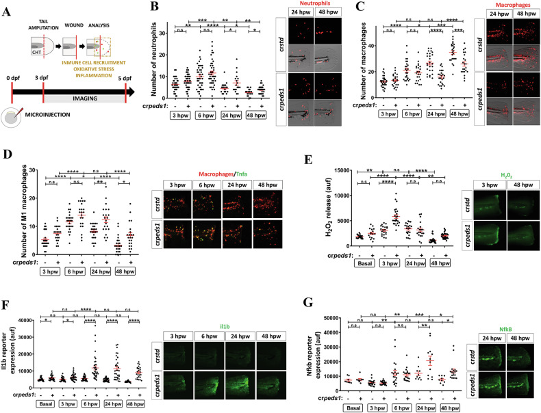
Fig. 3 Peds1 deficiency impairs inflammation resolution and causes aberrant immune cells recruitment in a sterile tail injury model.

A Schematic of the experimental procedure used for tail injury assays. One-cell stage zebrafish eggs were microinjected with std or peds1 crRNA/Cas9 complexes. At 3 dpf larvae were tail amputated and then inflammation and immune cell recruitment at the injury site (dotted lines) were analyzed at 3, 6, 24 and 48 h post-wounding (hpw) by fluorescence microscopy. The wound area is defined as the region between the amputation edge and the end of the caudal hematopoietic tissue (CHT). B, C Number of neutrophil and macrophages recruited at the injury site and representative merge images (brightfield and red channel; Tg(lyz:DsRED2)nz50 and Tg(mfap4.1:Tomato)xt12, respectively). D Number of M1-like macrophages (Tnfa+) recruited at the injury site and representative merge images (red channel: Tg(mfap4.1:Tomato)xt12 and green channel: Tg(tnfa:eGFP-F)ump5). E Analysis of H2O2 release at the wound site using the fluorogenic substrate acetyl-pentafluor-obenzene sulphonyl fluorescein. EG Analysis and representative images of Il1b (green channel; Tg(il1b:GFP-F)ump3) and Nfkb fluorescent reporter expression levels (green channel; Tg(NFkB-RE:eGFP)sh235) at the wound site. Each point represents one larva and the mean ± SEM of each group is also shown. P-values were calculated using one-way ANOVA and Tukey’s multiple range. n.s, not significant, *p ≤ 0.05, **p ≤ 0.01, ***p ≤ 0.001, ****p ≤ 0.001. auf arbitrary units of fluorescence.

Acknowledgments
This image is the copyrighted work of the attributed author or publisher, and ZFIN has permission only to display this image to its users. Additional permissions should be obtained from the applicable author or publisher of the image. Full text @ Cell Death Discov