|
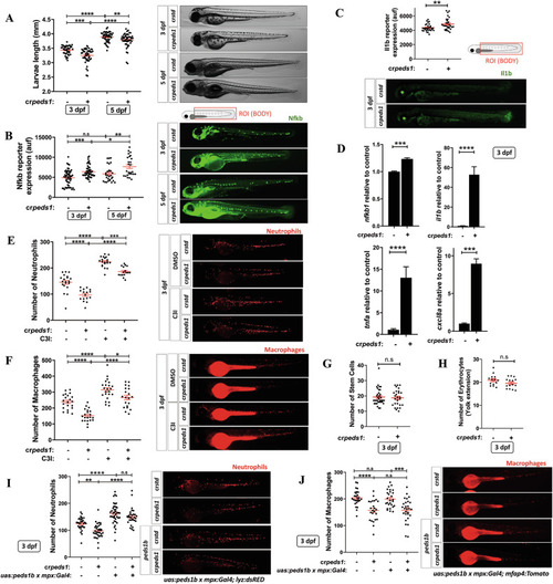
Peds1 deficiency promotes a proinflammatory stage with increased neutrophil and macrophage apoptosis. One-cell stage zebrafish eggs were microinjected with std or peds1 crRNA/Cas9 complexes and the following parameters were evaluated: (A) Larval length and representative images (brightfield channel) at 3- and 5 dpf, (B) Nfkb fluorescent reporter expression levels and representative images (green channel; Tg(NFkB-RE:eGFP)) at 3 and 5 dpf analyzed by fluorescence microscopy, (C) Il1b fluorescent reporter expression levels and representative images (green channel; Tg(il1b:GFP-F)ump3) at 3 dpf analyzed by fluorescence microscopy. D Transcript levels of the indicated genes analyzed at 3 dpf by RT-qPCR. E, F Number of neutrophils and macrophages and representative images of microinjected larvae after 48 h of treatment with caspase 3 inhibitor (C3I) or vehicle (DMSO) were quantitated by fluorescence microscopy (red channel; Tg(lyz:DsRED2) and Tg(mfap4.1:Tomato), respectively). G, H Hematopoietic stem and progenitor cells (green channel; Tg(-6.0itga2b:eGFP)) and erythrocytes (red channel: Tg(gata1a:dsRED)) number were quantitated by fluorescence microscopy. I, J Number of neutrophils and macrophages and representative images of larvae microinjected to specifically express peds1 in neutrophils using the neutrophil-specific myeloperoxidase (mpx) promoter and analyzed by fluorescence microscopy. Each point represents one larva and the mean ± SEM of each group is also shown. P-values were calculated using one-way ANOVA Tukey’s and multiple range or unpaired Student’s t-test. n.s, not significant, *p ≤ 0.05, **p ≤ 0.01, ***p ≤ 0.001, ****p ≤ 0.001. auf arbitrary units of fluorescence.
|