|
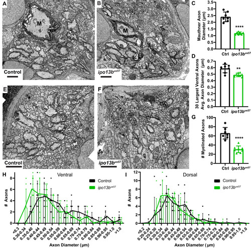
Electron microscopy of axon diameter growth defects in importin 13b mutants. A Representative electron micrographs of cross sections of the ventral spinal cord at 7 dpf in control and Bipo13bue57 animals. The Mauthner axon is labelled ‘M’. C Quantification of Mauthner axon diameter from 7 dpf electron micrographs (two-tailed unpaired t-test with Welch’s correction, n = 7 axons from individual animals, p < 0.0001). D Mean diameter for the 30 largest axons in each hemi ventral spinal cord at 7 dpf, excluding Mauthner (two-tailed unpaired t-test with Welch’s correction, n = 7 control and 8 ipo13bue57 animals, p = 0.006). E Representative electron micrographs of cross sections of the dorsal spinal cord at 7 dpf in control and Fipo13bue57 zebrafish. G Number of myelinated axons in the dorsal and ventral tracts per hemi spinal cord at 7 dpf (two-tailed unpaired t-test, n = 7 control and 8 ipo13bue57 animals, p < 0.0001). H Distribution of axon diameters for the 30 largest axons in each hemi ventral spinal cord at 7 dpf, excluding Mauthner (n = 7 control and 8 ipo13bue57 animals). I Distribution of axon diameters for the 30 largest axons in each hemi dorsal spinal cord at 7 dpf (n = 7 control and 8 ipo13bue57 animals). All data are presented as mean values ± SD. **p < 0.01, ****p < 0.0001. Scale bars = 1 µm. Source data are provided as a Source Data file.
|