|
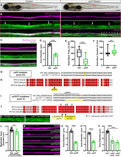
Identification of importin 13b mutants with reduced axon diameter growth. A Brightfield images of control and ue57 mutant zebrafish at 5 dpf, depicting normal growth and gross morphology. B The Mauthner axon (somite 15) labelled using Tg(hspGFF62A:Gal4); Tg(UAS:mRFP) in control (left panels) and ue57 mutant (right panels) is of smaller diameter in mutants at 5 dpf. Labelling of myelin with Tg(mbp:eGFP-CAAX) shows that the Mauthner axon is myelinated (white arrows). Magnification of the mutant Mauthner axon (region within the dashed rectangle) is shown in (C) with the myelin along the Mauthner axon outlined with dashed lines. D Mauthner axon diameter measured at somite 15 using the Tg(hspGFF62A:Gal4); Tg(UAS:mRFP) reporter at 5 dpf (two-tailed unpaired t-test, n = 10 axons from individual animals per genotype, p < 0.0001). E In a 20 min open field test at 5 dpf, ue57 mutant zebrafish initiate fewer swim bouts than controls; however, swim bouts are slightly longer in length (F) (two-tailed Mann–Whitney test, p < 0.0001 for (E) and p = 0.001 for (F), n = 144 control and 31 ue57 animals). G A region of exon 21 (last exon) of the ipo13b gene where a C > T base pair change (highlighted in red) was identified in ue57 mutants. H This base pair change results in the introduction of a premature stop codon in the highly conserved C-terminal region of importin 13b, predicted to result in a truncated protein missing the last 30 amino acids. I Overview of a region in exon 3 of the ipo13b gene indicating the site targeted with a sgRNA for Cas9-mediated DNA cleavage (PAM sequence highlighted in red), and the resulting mutations in the ue76 and ue77 mutant lines. J The mutations disrupt key residues previously shown to bind Ran-GTP (marked with #)57, and result in frame shifts followed by premature stop codons. K Representative images of the myelinated Mauthner axon (somite 15, 5 dpf) labelled using Tg(mbp:eGFP-CAAX) in control and ue57 zebrafish that were injected with 125 pg of ipo13b mRNA at the single cell stage, with quantification in (L) showing rescue of the axon diameter phenotype (two-tailed unpaired t-test, n = 15 control and 4 ue57 axons from individual animals, p = 0.096). M Representative images of the Mauthner axon (somite 15) labelled using the Tg(hspGFF62A:Gal4); Tg(UAS:GFP) reporter in 4–5 dpf control, ipo13bue76, ipo13bue77,ipo13bue57/ue77, ipo13bue57/ue77 zebrafish, with quantification of axon diameter compared to control siblings shown in (N–P) (N—two-tailed unpaired t-test, n = 6 axons from individual animals per genotype, p < 0.0001; O—two-tailed unpaired t-test, n = 7 axons from individual animals per genotype, p < 0.0001, P—One-way ANOVA with Tukey’s multiple comparisons test, n = 15 control, 6 ue57/ue76, and 8 ue57/ue77 axons from individual animals, p < 0.0001)). Data are presented as mean values ± SD, except E and F where data is presented as box and whisker plots with boxes indicating the median, 25th and 75th percentiles, and whiskers indicating the max and min. ***p < 0.001, ****p < 0.0001, ns = not significant. Scale bars: 300 µm (A), 20 µm (B), 10 µm (C, K, M). Source data are provided as a Source Data file.
|