FIGURE

Fig. 7

ID
ZDB-FIG-231208-41
Publication
Claude-Taupin et al., 2023 - The AMPK-Sirtuin 1-YAP axis is regulated by fluid flow intensity and controls autophagy flux in kidney epithelial cells
Other Figures
All Figure Page
Back to All Figure Page
Fig. 7

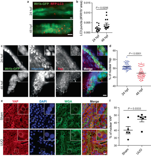
YAP subcellular localization is associated to autophagy activity in vivo.

a, b Representative images (a) and quantification (b) of RFP-LC3 puncta in Wt1b-GFP+ pronephros, at 24 h post fertilization (hpf) and 48 hpf. Data show the mean ± s.e.m.; n = 18 individual data points from 3 independent experiments, two-sided t-test. Scale bars, 10 µm. c, d Representative images (c) and quantification (d) of YAP nuclear localization in Wt1b-GFP+ pronephros at 24 and 48 hpf. Nucleus were labeled with Hoechst. Data show the mean ± s.e.m.; n = 60 individual data points from 3 independent experiments, two-sided t-test. Scale bars, 10 µm. e, f Mice subjected to unilateral ureteral obstruction (UUO) or sham operation were euthanized 24 h after surgery and kidney sections subjected to immunohistochemistry for YAP and renal tubule marker Wheat Germ Agglutinin (WGA). e Representative images. Scale bars, 10 µm. f Quantification of YAP nuclear levels in the renal tubules (WGA+). Data show the mean ± s.e.m.; n = 5 (sham) and n = 6 (UUO) different mice. two-sided t-test. Source data are provided as a Source Data file.

Expression Data

Expression Detail
Antibody Labeling
Phenotype Data

Phenotype Detail
Acknowledgments
This image is the copyrighted work of the attributed author or publisher, and ZFIN has permission only to display this image to its users. Additional permissions should be obtained from the applicable author or publisher of the image. Full text @ Nat. Commun.