|
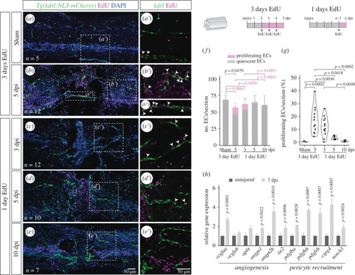
Endothelial proliferation during vascular repair. (a–e) Longitudinal SC sections with labelled ECs (Tg(kdrl:NLS-mCherry)), EdU staining and nuclear DAPI staining, with magnified views (a′–e′) showing proliferating ECs (arrowheads). Repeated EdU injections were performed three consecutive days before SC collection at 5 days after sham/spinal cord contusion injury (a,b). Single EdU injections were administered 1 day before SC collection at 3, 5 and 10 dpi (c,d,e). (f) Total number of ECs per section, grouped as proliferating (EdU+) or quiescent (EdU−) cells. (g) Fraction of proliferating ECs (EdU+ ECs / total ECs) per section. (h) Relative gene expression in uninjured (n = 6) and 7 dpi (n = 2) SCs, measured by qPCR, standardized to gapdh and normalized to the uninjured mean. Data represent mean ± s.d. Statistical tests: Kruskal–Wallis test followed by Dunn's multiple comparisons post hoc test (f,g); two-tailed unpaired t-test (O); (p-values > 0.05 are not shown).
|