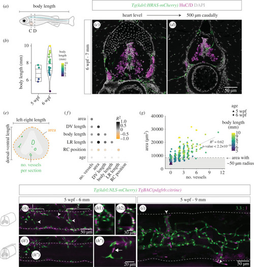
Spinal cord vascularization in juvenile zebrafish. (a) Definition of body length and rostral–caudal region analysed in juvenile zebrafish. (b) Body length (y-axis and symbol colour) in juvenile fish with 5 (n = 4) or 6 (n = 20) weeks post fertilization (wpf). (c,d) 6 wpf fish sections at the heart level (c) and 500 µm caudally (d) with labelled endothelial cells (Tg(kdrl:HRAS-mCherry)), HuC/D+ neurons and nuclear DAPI staining. (e) Schematic showing the parameters quantified in the imaged sections. (f) Correlation matrix dot plot of the quantified parameters: area; DV (dorsal–ventral) length; body length; LR (left-right) length; RC (rostral–caudal) position (starting at the heart level); age and number of vessels per section. Dot size and colour represent the Spearman correlation coefficient. (g) SC area relative to number of vessels per section in all sections analysed. Symbol shape indicates age and symbol colour indicates body length. Dashed line corresponds to average SC area in sections with 1 vessel inside. (j) 5 wpf juvenile with 6 mm with labelled endothelial nuclei (Tg(kdrl:NLS-mCherry)) and perivascular cells (TgBAC(pdgfrb:citrine)) (arrowheads identify the perineural vascular plexus and the asterisk marks a vessel inside the spinal cord). (h1,h2) Transversal views of the regions marked with brackets in h. (h′) Ingressing blood vessel in the central region with co-recruited mural cells (arrow) (magnification in h″). (i) A 9 mm fish SC with a more complex vasculature and high perivascular coverage (3.3 ECs per perivascular cell ±0.28, n = 3). Arrowheads label points of connection between the intraspinal network and external vessels.
|

