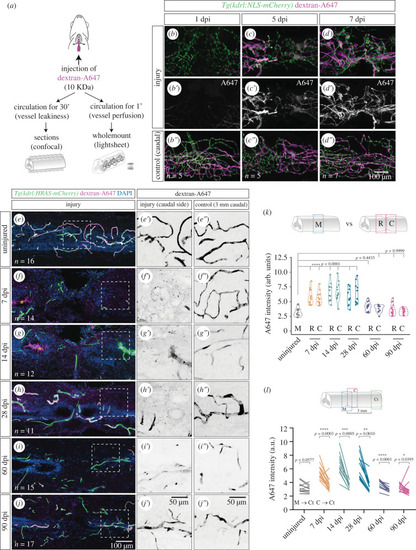
Re-establishment of the blood-spinal cord barrier after injury. (a) Schematic of the vessel perfusion/permeability assay with 10KDa dextran-Alexa647 (A647), adapted from [37]. (b–d″). Wholemount SCs with labelled endothelial nuclei (Tg(kdrl:NLS-mCherry)), perfused with dextran-A647 for 1 min and analysed at 1, 5 and 7 dpi. (b′–d′). A region 2 mm caudally to the injury is used as control of the injection (b″–d″). (e–j) Longitudinal sections of contusion-injured SCs with labelled ECs (Tg(kdrl:HRAS-mCherry)), perfused with dextran-A647 for 30 min and with DAPI-stained nuclei. Orange asterisks highlight the accumulation of dextran-A647 in the central canal in f,g. Magnified views of the dextran-A647 signal in the caudal side of the injury are shown in e′–j′. and a control region 3 mm caudal to the injury is shown in e″–j″. (k) Quantification of extravascular intensity of dextran-A647 in the rostral (R) and caudal (C) side of the injury and compared to the middle region (M) of uninjured SCs. (l) Comparison of dextran-A647 extravascular intensity in the caudal side of the injury (or a corresponding region in uninjured SCs) with a control region 3 mm caudal to the injury. Each line shows the change between the two regions in individual SCs. Statistical tests: Kruskal–Wallis test followed by Dunn's multiple comparisons post hoc test relative to uninjured control (k) and Wilcoxon matched-pairs signed rank test (l).
|
