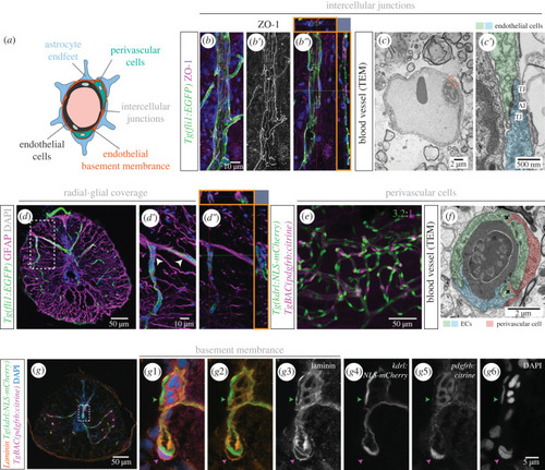
Organization of the blood-spinal cord barrier in adult zebrafish. (a) Schematic of BSCB components. (b-b″). Tg(fli1:EGFP) SC section, co-stained with the tight junction marker ZO-1 (b′). Orthogonal views (orange squares in b″) shows close proximity between ZO-1 and fli1:EGFP. (c-c′). TEM image of a blood vessel, with a magnification of the orange box in (c′) showing intercellular junctions between ECs: tight junctions (TJ) and adherens junctions (AJ). (d–d″). Transverse SC section labelled ECs Tg(fli1:EGFP) and radial glial cells (anti-GFAP antibody) with projections adjacent to ECs (arrowheads in (d′) and orthogonal views in orange squares in (d″)). (e) Wholemount SC with labelled endothelial nuclei (Tg(kdrl:NLS-mCherry)) and pericytes (TgBAC(pdgfrb:citrine)) (3.2 ECs per perivascular cell ± 0.59, n = 7). (f) TEM image of a cross-section of a blood vessel, showing two contacting ECs (in blue and green) and an associated pericyte (in red). (g) Transverse SC section with labelled endothelial nuclei (Tg(kdrl:NLS-mCherry)), pericytes (TgBAC(pdgfrb:citrine)), co-labelled with an anti-laminin antibody to identify the basement membrane (BM) and DAPI-labelled nuclei. (g1–g6) Magnifications of the region labelled in (g) showing the separate channels. The green arrowhead points to the BM over an endothelial nucleus and the magenta arrowhead identifies a pericyte embedded in the endothelial BM.
|
