FIGURE

Figure 7

ID
ZDB-FIG-230228-180
Publication
Le Mentec et al., 2023 - A New In Vivo Zebrafish Bioassay Evaluating Liver Steatosis Identifies DDE as a Steatogenic Endocrine Disruptor, Partly through SCD1 Regulation
Other Figures
All Figure Page
Back to All Figure Page
Figure 7

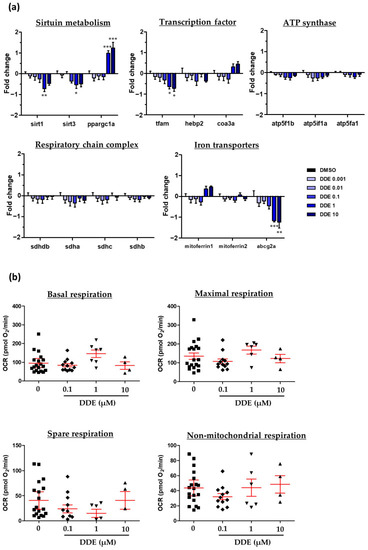
Assessment of potential mitochondrial dysfunction induced by DDE. Zebrafish larvae at 3 dpf were exposed to DDE (from 1 nM to 10 µM) or to control vehicle during 48 h. (a) Impact of DDE on the mRNA expression of several genes involved in mitochondrial metabolism. mRNA expression was evaluated by RT-qPCR gene expression analysis by using the microfluidic Fluidigm-Biomark technology. Data are expressed relative to mRNA levels measured in DMSO control larvae, set at 0 (log 2 fold change) (n ≥ 8). (b) Evaluation of respiration after DDE exposure of zebrafish larvae. Measurements on zebrafish larvae were realized on a Seahorse XFe24 Analyzer of mitochondrial oxygen consumption. Values are the mean of oxygen consumption rate (OCR, in pmol of O2/min) ± SEM measured from at least four larvae per condition. Values are the mean ± SEM. * p < 0.05, ** p < 0.01, *** p < 0.001, by comparison with control group using ANOVA and Dunnett’s test.

Expression Data

Expression Detail
Antibody Labeling
Phenotype Data

Phenotype Detail
Acknowledgments
This image is the copyrighted work of the attributed author or publisher, and ZFIN has permission only to display this image to its users. Additional permissions should be obtained from the applicable author or publisher of the image. Full text @ Int. J. Mol. Sci.