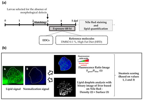
Experimental design of the StAZ bioassay (a) Steatogenic Assay on Zebrafish (StAZ) protocol. Zebrafish larvae at 3 days post-fertilization (dpf) were exposed to the selected compounds at non-toxic concentrations, to the positive control (HFD), or to vehicle alone (DMSO 0.1%), during 48 h. Following exposure, larvae were euthanized and fixed, and hepatic lipid accumulation was measured using Nile red staining and fluorescence signal quantification. (b) Fluorescent signal quantification workflow. After image acquisition of stained zebrafish larvae with confocal fluorescence microscopy, two types of images were obtained: a first one with a green signal—characteristic of neutral lipid fluorescence, and a second one with a blue signal for normalization—insensitive to neutral lipids (images A and B, respectively). Using Fiji imaging processing software and home-made macros, three parameters were calculated: (1) the ratio of fluorescence intensity of image A to image B per liver area (F green/F blue), (2) the amount of lipid droplets per liver area, and (3) the surface occupied by lipid droplets per liver area. Based on these three parameters, a steatosis score was calculated. Each parameter was pondered by a specific coefficient determined empirically.
|
