
Figure EV5
- ID
- ZDB-FIG-220131-583
- Publication
- Wu et al., 2021 - Sensing of mycobacterial arabinogalactan by galectin-9 exacerbates mycobacterial infection
- Other Figures
- All Figure Page
- Back to All Figure Page
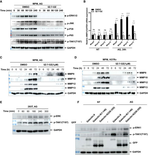
Immunoblots of cell lysates of peritoneal macrophages stimulated with AG (1 μg/ml) in the absence or presence of TAK1 inhibitor 5Z‐7‐OZ (1 μM) for indicated times. Data are representative of qPCR analysis of Immunoblots of cell supernatants to analyze secreted MMP9, MMP10, MMP12, and MMP13 by mouse peritoneal macrophages stimulated with AG (1 μg/ml) for indicated times in the absence or presence of TAK1 inhibitor 5Z‐7‐OZ (1 μM); GADPH of cell lysates served as the loading control. Immunoblots of cell supernatants to analyze secreted MMP9, MMP10, MMP12, and MMP13 by mouse peritoneal macrophages infected with H37Rv for indicated times (MOI = 5) in the absence or presence of TAK1 inhibitor 5Z‐7‐OZ (1 μM); GADPH of cell lysates served as the loading control. Immunoblots of cell lysates of HEK293T cells stimulated with AG (1 μg/ml) for the indicated time to analyze p‐ERK1/2 and p‐TAK1(T187). GADPH of cell lysates is shown as loading control. Immunoblots of lysates of HEK293T cells stimulated with AG (1 μg/ml) for 3 h after transfection of the indicated plasmids for 48 h. Data information: Data in (B) are means ± SD averaged from 3 independent experiments performed with technical triplicates and each symbol represents the mean of technical triplicates. Two‐way ANOVA followed by Dunnett's |