|
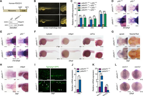
<italic>Pdcd11</italic> mutant zebrafish favor inflammatory macrophage generation.a Schematic diagram showing the two major function domains of PDCD11: N-terminal Ribosome domain (1–1407) and C-Terminal Coil domain (1408–1871). b Gross morphology of 3 dpf WT and pdcd11 mutants. c qPCR confirmation of hyperactivated inflammatory pathway genes, including tnfa, il1b, and il6, in 22 hpf pdcd11 mutants, which could not be restored by combined p53 mutation. d–eIl6 and il1b expression in the brain of WT or pdcd11 mutants with p53 mutated or not examined by WISH at 52 hpf. f WISH analysis of l-plastin, mfap4, and csf1ra expression in 24 hpf WT and pdcd11 mutant embryos. Red arrows indicate csf1ra-positive cells in the brain. g Microglia development in 3 dpf WT and pdcd11 mutants by WISH assessment of apoeb and Neutral Red staining. hL-plastin and mfap4 expression pattern in 3 dpf WT and pdcd11 mutants. i Morphology of macrophages in the brain and caudal hematopoietic tissue (CHT) were examined using the Tg(mpeg1:GFP) transgenic line. White arrows indicate the vacuolated macrophages found in the pdcd11 mutant. j Increased tnfa mRNA expression in sorted macrophages Tg(mpeg1:GFP) from 60 hpf pdcd11 mutants as compared with WT controls. k qPCR examination of the expression of macrophage-related genes cxcr3.1 and cxcr3.2 in 22 hpf WT and pdcd11 mutants. l WISH analysis of cxcr3.1 and cxcr3.2 expression in 22 hpf WT and pdcd11 mutants. The number positioned in the lower right corner of d–h, l represent the number of zebrafish embryos shown positive phenotypes versus the total number of embryos examined. Scale bar: 100 μm. Black horizontal lines indicate mean ± standard error of mean (SEM). Means ± SEM are shown for three independent experiments. *P < 0.05; **P < 0.01; ***P < 0.001 (Student’s t test).
|

