|
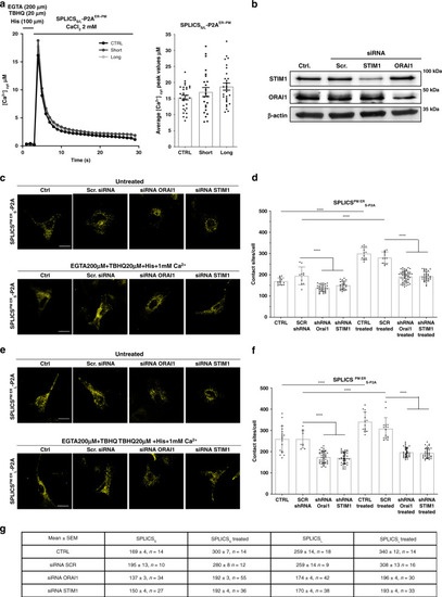
Deep proof-of-concept characterization of the ER–PM reporter.a Cytosolic Ca2+ transients (left) and quantification (right) upon induction of the ER Ca2+ depletion in cells overexpressing SPLICSS-P2AER–PM and SPLICSL-P2AER–PM probes. To induce the ER Ca2+ depletion cells were treated with 20 µM 2,5-tertbutylhydroquinone (THBQ), 100 μM histamine, and 200 μM EGTA. Where indicated 2 mM CaCl2 was then applied to induce Ca2+ influx. The traces are the media of at least 15 independent measurements obtained from two independent transfections, mean ± SEM. b Expression levels of STIM1 and ORAI1 proteins in mock (Ctrl), siRNA scramble or siRNA STIM1, or SiRNA ORAI1 treated cells were analyzed by Western blotting with an anti-STIM1 or anti-ORAI1 antibodies. Equal amount of total loaded lysate was verified by incubation with anti-β actin antibody. Effect of STIM1/ORAI1 silencing and the ER Ca2+ depletion on short c and long e range contacts between the ER and PM. Representative microscope single plane images of HeLa cells expressing the SPLICSS/L-P2AER–PM untreated (top) or treated for 5 min (bottom) with 200 μM EGTA, 20 µM THBQ, 100 μM histamine, and then for additional 5 min with 1 mM CaCl2 supplemented to KRB. Scale bar 25 µm. Quantification of SPLICSS-P2AER–PMd and SPLICSL-P2AER–PMf contacts by 3D rendering of complete Z-stacks, mean ± SEM. The data were obtained from three independent transfections (****p ≤ 0.0001 unpaired two-tailed t-test). g Mean ± SEM values of the number of PM–ER contacts in control conditions, upon SOCE activation and upon siRNA scramble or siRNA STIM1 or SiRNA ORAI1 incubation. Source data are provided as a source data file.
|